38 drag the labels onto the diagram of the cns meninges.
This stimulates the visual centres in the brain, giving us the sensation of seeing. In this interactive, you can label parts of the human eye. Use your mouse or finger to hover over a box to highlight the part to be named. Drag and drop the text labels onto the boxes next to the eye diagram. Brain Label (Remote) Shannan Muskopf December 29, 2020. This brain labeling activity was created for remote learners as an alternative to the labeling and coloring worksheet we would traditionally do in class. Instead of coloring and labeling on printouts, students use google slides to drag labels to the images or type the answers into text boxes.
We review their content and use your feedback to keep the quality high. 100% (11 ratings) Answer The label is indicated from RIGHT SIDE of image to …. View the full answer. Transcribed image text: Part A Drag the labels onto the diagram to identify the spinal nerve roots and meninges Reset Help Ventral Pia mater Meninges Dorsal root Dura mater.
Drag the labels onto the diagram of the cns meninges.
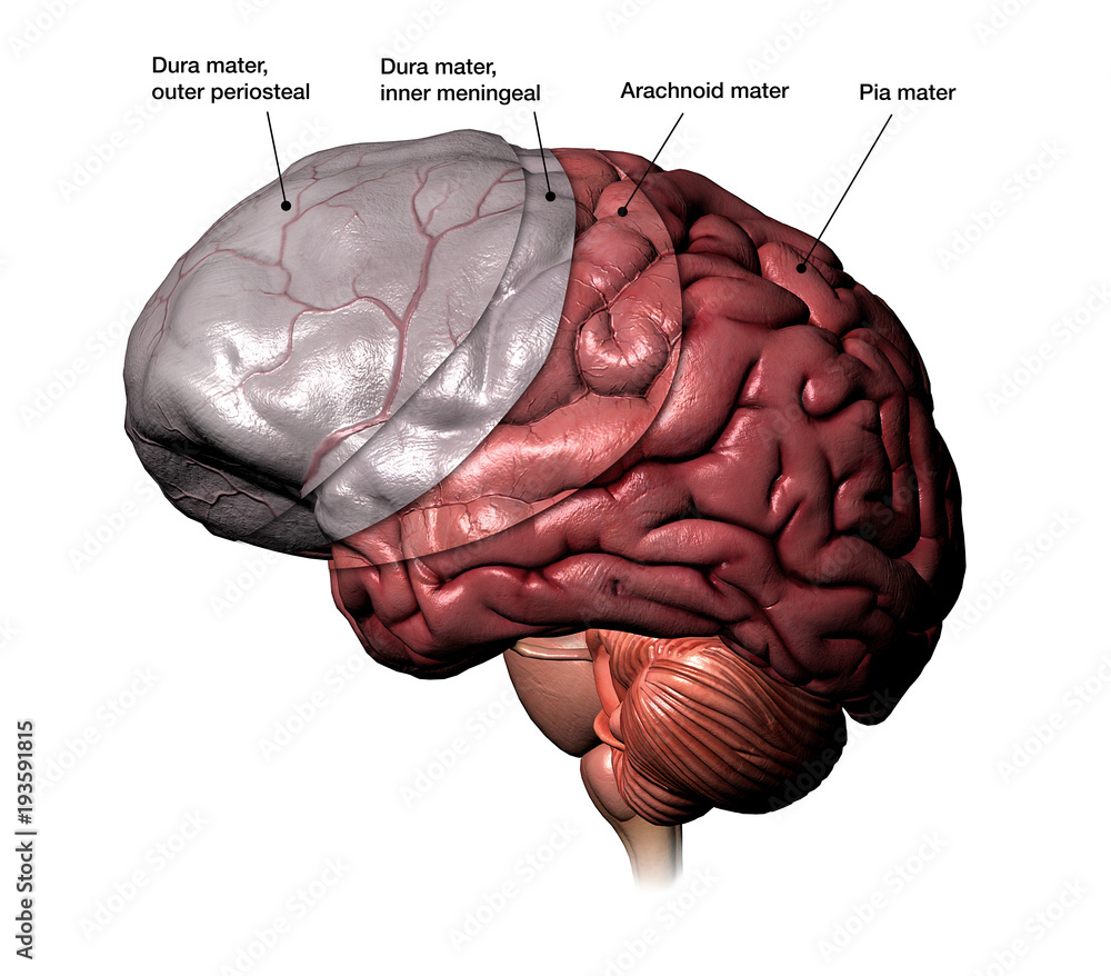
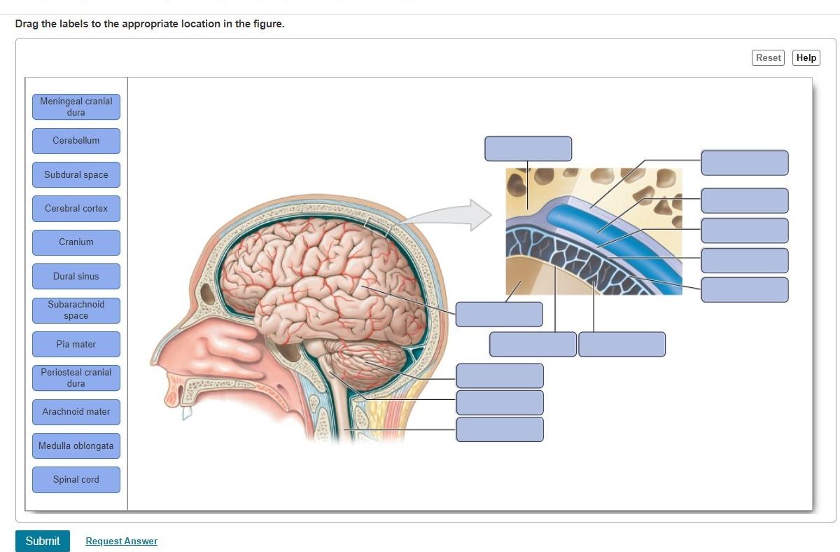
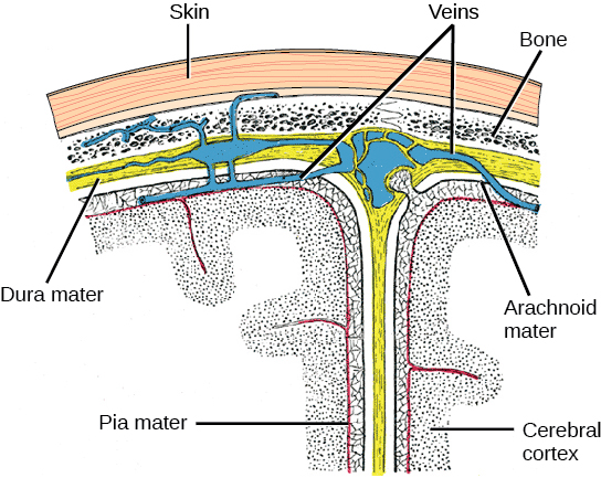
The meninges is a layered unit of membranous connective tissue that covers the brain and spinal cord.These coverings encase central nervous system structures so that they are not in direct contact with the bones of the spinal column or skull. The meninges are composed of three membrane layers known as the dura mater, arachnoid mater, and pia mater. Art-labeling Activities. This activity contains 3 questions. Label the regions on the diencephalon and brain stem (posterior view). For each item below, use the pull-down menu to select the letter that labels the correct part of the image. Match the following labels to the proper locations on the sagittal section of the brain. The meninges are three connective tissue membranes that lie just external to the brain. The function of theses layers are to: 1) cover and protect the brain, 2) protect blood vessels and enclose venous sinuses, 3) contain cerebral spinal fluid, and 4) form partitions within the skull.
Drag the labels onto the diagram of the cns meninges.. View Screen Shot 2019-02-05 at 8.26.00 PM.png from BSC 2086L at University of South Florida. Drag the labels onto the diagram to identify the cranial meninges and associated structures. Drag the labels onto the diagram to identify the spinal nerve roots and meninges Drag the labels onto the diagram to identify the gross anatomical structures of the spinal cord. Drag the labels onto the diagram to identify the parts of the spinal cord (transverse section, showing white matter). Spinal Cord Anatomy. In adults, the spinal cord is usually 40cm long and 2cm wide. It forms a vital link between the brain and the body. The spinal cord is divided into five different parts. Several spinal nerves emerge out of each segment of the spinal cord. There are 8 pairs of cervical, 5 lumbar, 12 thoracics, 5 sacral and 1 coccygeal pair ... •Cranial meninges •Dura mater, arachnoid mater, and pia mater •Cerebrospinal fluid •Provides protection of the brain and spinal cord •Provides support •Transports nutrients to the CNS tissue •Transports waste away from the CNS •Blood-brain barrier •Maintains a constant environment, necessary for both control
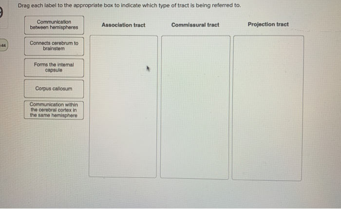
Meninges of the Spinal Cord and Brain are similar Dura Mater -outermost membrane of tough collagen fibers -epidural space between the dura mater and the vertebral canal is filled with fat and blood vessels •epidural anesthesia is delivered into the epidural space • Arachnoid (Mater) -middle layer composed of a simple squamous epithelium HW 7 Due: 11:59pm on Friday, October 27, 2017 To understand how points are awarded, read the Grading Policy for this assignment. Art-labeling Activity: Organization of the Nervous System Learning Goal: To learn the divisions and receptors of the nervous system. Label the divisions and receptors of the nervous system. Part A Drag the labels onto the diagram to identify the divisions and ... Cns central nervous system 7. Drag the labels onto the diagram to identify parts of the neuromuscular junction. What part of the nervous system performs information processing and integration. Drag the labels onto the diagram to identify the components of somatic sensory pathways. By antlab plays quiz not verified by sporcle. Sheep Brain Dissection Guide Now onto the Dissection Proper. ... The meninges are the protective coverings, which enclose the brain and spinal cord. The dura mater, the tough outer layer, will have been mostly ... the central nervous system). This is the corpus callosum. It is so big that different parts of it get different names.
Drag the labels onto the diagram to identify structural features associated with skeletal muscle. The structure indicated by label e is part of which of the following. Drag the labels onto the diagram to identify the various muscle structures. Learn vocabulary terms and more with flashcards games and other study tools. Drag the labels onto the diagram to identify steps in response to low blood pressure. Who are the experts? Experts are tested by Chegg as specialists in their subject area. We review their content and use your feedback to keep the quality high. The above diagram is filled with all the appropriate terms . The above diagram represent the adaption ... Psychology Nervous System Diagram Worksheets/Notebook Pages. by. Green Eyes Learning. $4.00. PDF. This 5 page PDF provides you with 3 clean & organized diagrams for students to label. These diagrams include the neuron, brain, & nervous system. Subjects: Anatomy, Biology, Psychology. Drag the labels onto the diagram to identify the cranial meninges and associated structures. look at pic Drag the labels to identify the landmarks and features on one of the cerebral hemispheres.
Look no further than these interactive, exam-style anatomy quizzes. Learn anatomy faster and. remember everything you learn. Start Now. <. General Structure of a Neuron (Nerve Cell) >. Nose and Nasal Cavity: Openings and Support Structures.
Drag the labels to identify the structures of a long bone. If an expanded label shows a list of structures, it is a group label. Drag the appropriate labels to their respective targets. Part a drag the labels onto the diagram to identify the structures associated with ganglia in sympathetic pathways (collateral ganglia) reset help lateralam .
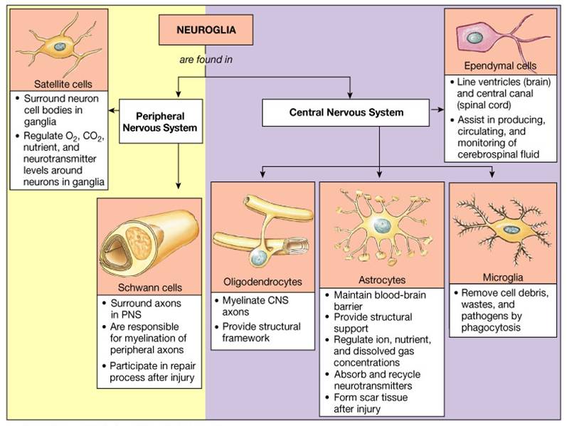
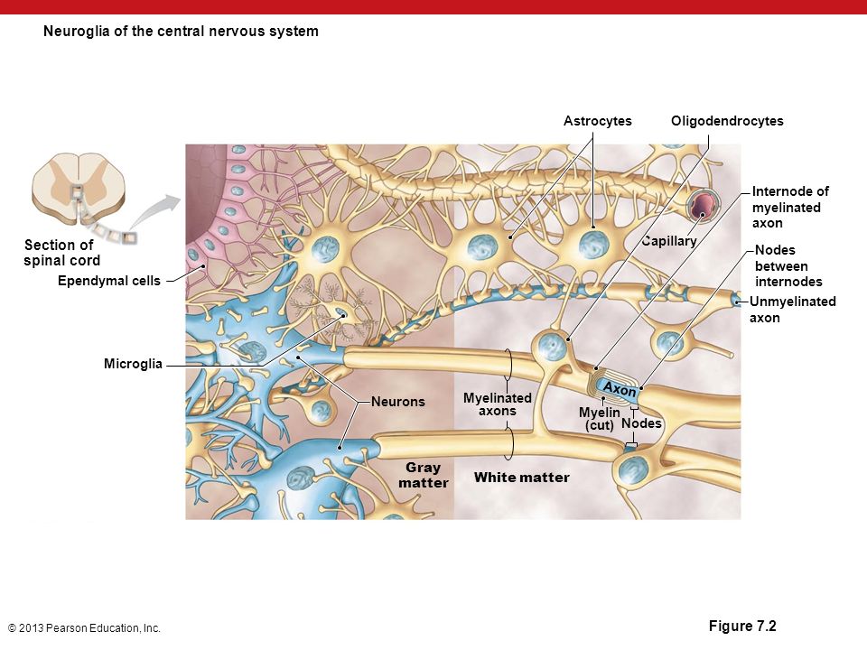
Drag the labels onto the diagram to identify the gross anatomical structures of the spinal cord. look at pic. Drag the labels onto the diagram to identify the spinal nerve roots and meninges. look at pic. ... while Central Nervous System (CNS) neuroglial cells called _____ are responsible for the formation of a myelin sheath. Schwaan cells ...
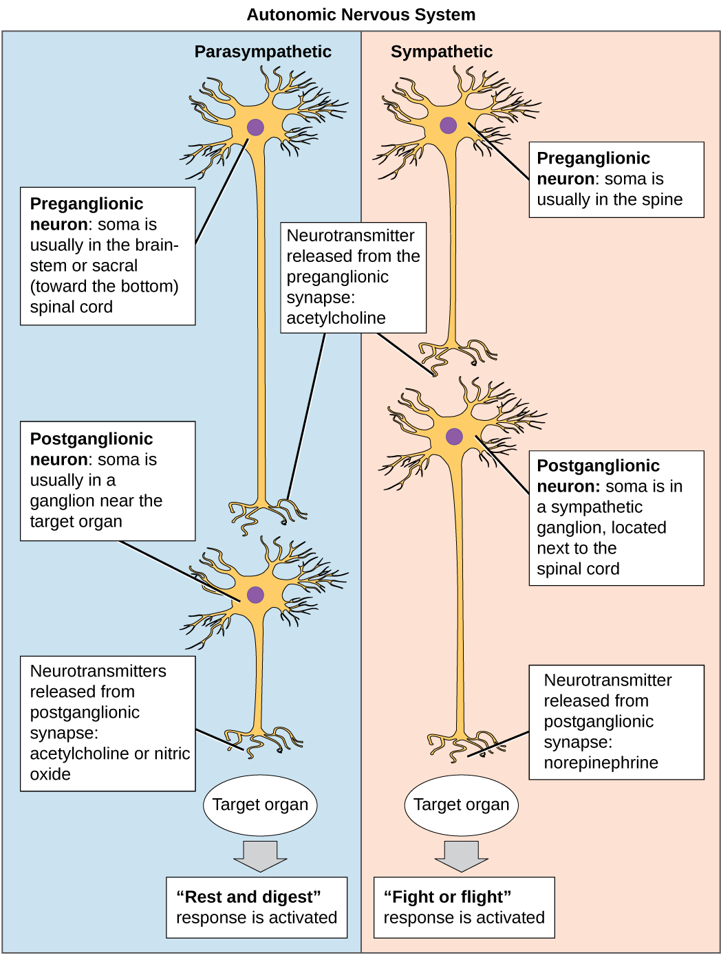
Drag the labels onto the diagram of neurochemical communication at an autonomic synapse. ANSWER: Correct. Art-labeling Activity Figure 11. Label the parts of the neuromuscular junction. Part A. Drag the labels onto the diagram to identify parts of the neuromuscular junction. ANSWER: Reset Help. Action potential arrives at varicosity.
Drag the labels onto the diagram to identify the parts of the spinal cord (transverse section, showing gray matter). 1. central canal. 2. anterior gray commissure. 3. anterior median fissure. 4. posterior median sulcus. 5. posterior gray horn. 6. lateral gray horn. 7. dorsal root. 8. anterior gray horn.
41 drag the labels onto the diagram to identify factors that affect mean arterial pressure. Written By Chelsea P. Mariano. Friday, November 26, 2021 Add Comment Edit. Terms in this set (33) Drag the labels onto the diagram to identify aspects of gas transport and exchange.

For convenience, the nervous system, is considered in terms of two principal divisions: the central nervous system and the peripheral nervous system. The central nervous system (CNS) consists of the brain and spinal cord, which primarily interpret incoming sensory informa-tion and issue instructions based on that information and on past experience.
The brain and spinal cord are enveloped within three layers of membrane collectively known as the meninges, with the cranial meninges specifically referring to the section that covers the brain. From superficial to deep, the three layers are the dura, arachnoid, and pia—the term "mater," Latin for mother, often follows these names (i.e., dura mater, arachnoid mater, pia mater).[1]
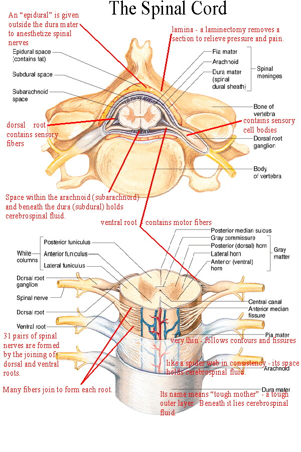
Figure 14.2a The Spinal Cord and Spinal Meninges Anterior view of spinal cord showing meninges and spinal nerves. For this view, the dura and arachnoid membranes have been cut longitudinally and retracted (pulled aside); notice the blood vessels that run in the subarachnoid space, bound to the outer surface of the delicate pia mater.
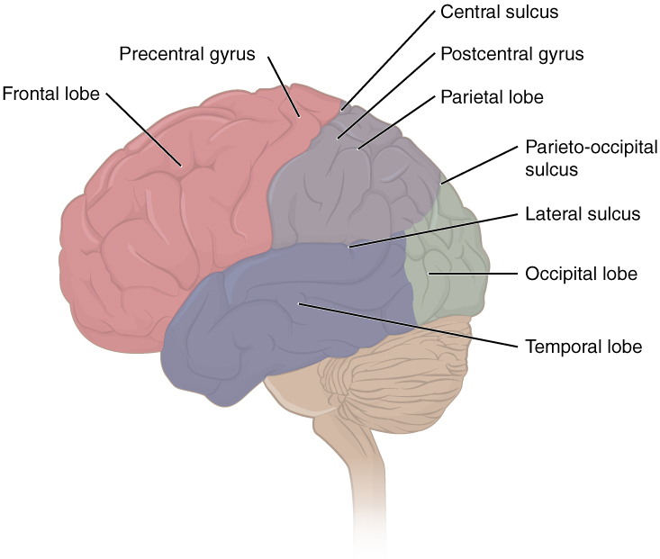
Drag the labels onto the diagram to identify the cranial meninges and associated structures. look at pic Drag the labels to identify the landmarks and features on one of the cerebral hemispheres.

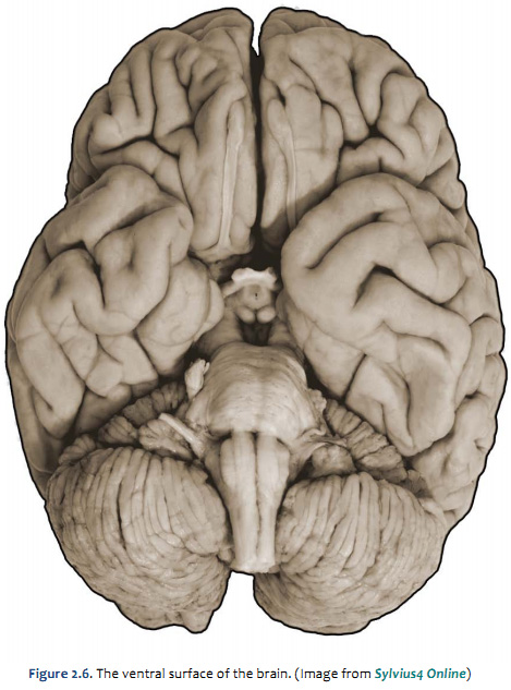
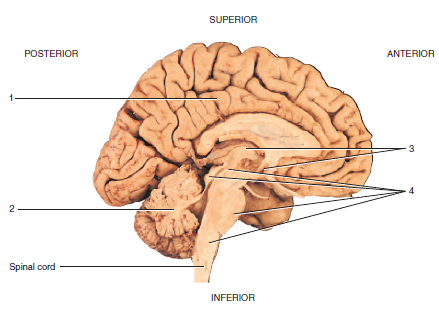
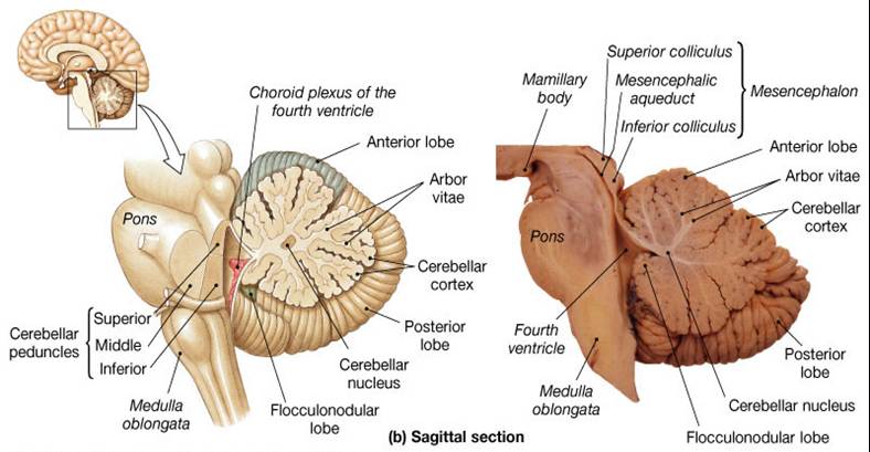
The brain and the spinal cord are the central nervous system, and they represent the main organs of the nervous system. The spinal cord is a single structure, whereas the adult brain is described in terms of four major regions: the cerebrum, the diencephalon, the brain stem, and the cerebellum. A person's conscious experiences are based on ...

The meninges are three connective tissue membranes that lie just external to the brain. The function of theses layers are to: 1) cover and protect the brain, 2) protect blood vessels and enclose venous sinuses, 3) contain cerebral spinal fluid, and 4) form partitions within the skull.

Art-labeling Activities. This activity contains 3 questions. Label the regions on the diencephalon and brain stem (posterior view). For each item below, use the pull-down menu to select the letter that labels the correct part of the image. Match the following labels to the proper locations on the sagittal section of the brain.
The meninges is a layered unit of membranous connective tissue that covers the brain and spinal cord.These coverings encase central nervous system structures so that they are not in direct contact with the bones of the spinal column or skull. The meninges are composed of three membrane layers known as the dura mater, arachnoid mater, and pia mater.


0 Response to "38 drag the labels onto the diagram of the cns meninges."
Post a Comment