37 inferior vena cava diagram
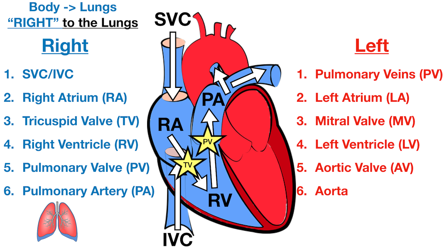
November 13, 2017 - Blood enters the heart through two large veins, the inferior and superior vena cava, emptying oxygen-poor blood from the body into the right atrium of the heart. After the kidneys have performed their cleansing function, the filtered, deoxygenated blood leaves the kidneys through the renal vein, moves up the inferior vena cava, and returns to the heart ...
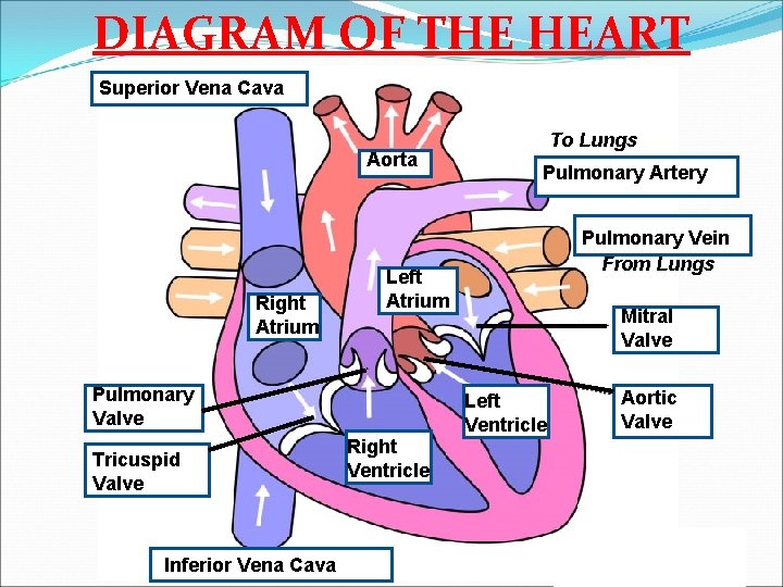
Superior vena cava Ascending aorta Right pulmonary artery Pulmonary trunk Right pulmonary veins Auricle of right atrium Right coronary artery (in atrioventricular sulcus) Right ventricle Inferior vena cava (a) Anterior view of the external heart C' 2019 Pearson Education. Aort'c arch Ligamentum arteriosum Left pulmonary artery Left pulmonary ve ns
Inferior vena cava diagram
6 Inferior Vena Cava. The great vessels of the heart function to carry blood to and from the heart as it pumps, located largely within the middle mediastinum. In this article we will consider the structure and anatomical relationships of the aorta, pulmonary arteries and veins, and the superior and inferior vena cavae. August 5, 2016 - Die obere und untere Hohlvene (Vena cava superior, Vena cava inferior) sind die zwei größten Venen im menschlichen Körper. Sie sammeln sauerstoffarmes Blut aus der Körperperipherie und leiten es zurück zum Herzen, genauer gesagt in den rechten Herzvorhof. Step 1 involves the superior vena cava (SVC) and inferior vena cava (IVC). They are the main blood vessels that carry the deoxygenated venous blood from the rest of the body to the right side of the heart, specifically the right atrium. The superior vena cava is located superiorly, and it carries the deoxygenated venous blood from the upper ...
Inferior vena cava diagram. The inferior vena cava (also known as IVC or the posterior vena cava) is a large vein that carries blood from the torso and lower body to the right side of the heart. From there the blood is pumped to the lungs to get oxygen before going to the left side of the heart to be pumped back out to the body. The IVC gets its name from its structure ... Recall that blood returning from the systemic circuit enters the right atrium (Figure 2) via the superior and inferior venae cavae and the coronary sinus, which drains the blood supply of the heart muscle. These vessels will be described more fully later in this section. The inferior vena cava (IVC) is the largest vein of the human body. It is located at the posterior abdominal wall on the right side of the aorta. The IVC's function is to carry the venous blood from the lower limbs and abdominopelvic region to the heart.. The inferior vena cava anatomy is essential due to the vein's great drainage area, which also makes it a hot topic for anatomy exams. untere/obere Hohlvene · Sie möchten laufend informiert werden? Newsletter abonnieren
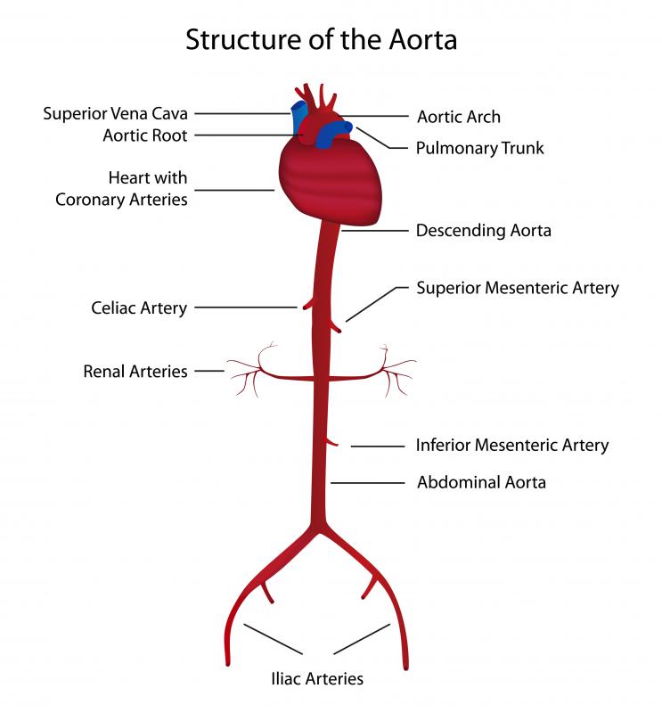
Vena Cava And Aorta Diagram. In this image, you will find phrenic artery, spleen, splenic artery, left renal artery, superior mesenteric artery, inferior mesenteric artery, left internal iliac artery in it. You may also find right external iliac artery, right common iliac artery, aorta, gonadal, right renal artery, common hepatic artery, left ... Superior Vena Cava & Inferior Vena Cava. The vena cava is the largest vein in the body that delivers oxygen-poor or deoxygenated blood to the right atrium of the heart. The superior vena cava comes from the upper part of the body, including the brain and arms, while the inferior vena cava comes from the abdominal area and legs. The inferior vena cava is a large vein that carries the deoxygenated blood from the lower and middle body into the right atrium of the heart.Drains to: Right atriumLatin: vena cava inferiorArtery: abdominal aortaSource: common iliac vein; lumbar veins; testic...Structure · Clinical significance Die Vena cava inferior entsteht aus dem Zusammenfluss der beiden Venae iliacae communes im Lendenbereich und zieht in Richtung Zwerchfell. Sie tritt durch das Hohlvenenloch (Foramen venae cavae) durch das Zwerchfell hindurch und verläuft in einer eigenen Pleurafalte (Plica venae cavae) zum ...
The pathway of blood flow through the heart begins as blood comes from the body and enters the heart through the superior and inferior vena cava indicated by the yellow star in the diagram below. Superior and inferior vena cavae and the coronary sinus 2. The right atrium receives deoxygenated blood through the superior and inferior vena cavas ... September 11, 2015 - Anomalien der Vena cava inferior sind angeborene Lage- und Verlaufsvarianten der unteren Hohlvene, die klinisch ... An inferior vena cava (IVC) filter is a small device that can stop blood clots from going up into the lungs. The inferior vena cava is a large vein in the middle of your body. The device is put in during a short surgery. Veins are the blood vessels that bring oxygen-poor blood and waste products back to the heart. 3. Superior Vena Caval 4. Inferior Vena Caval: Variations of ASD. Atrial Septal Defects are divided into three different types on the basis of the position of the hole (or holes) in the atrial septum. The first type of ASD is known as ostium primum defect, or simply, primum (number 1 in the diagram).
Journal Of Anatomy Absence At The Meeting Of The Anatomical Society References 1 Ernst N P A Case Of Congenital Atresia Of The Duodenum Treatedsuccessfully By Operation Brit Med Journal
February 24, 2020 - The inferior vena cava is also referred to as the posterior vena cava. The inferior vena cava is a large vein that carries de-oxygenated blood from the lower body to the heart.

Pengertian Vena Cava Superior Dan Inferior Sekolah007
Download scientific diagram | | Anatomy of major abdominal veins. Inferior vena cava segments adapted from Ref. (8). from publication: Thrombosis of the Abdominal Veins in Childhood | Abdominal venous thrombosis is a rare form of venous thromboembolic disease in children.
1
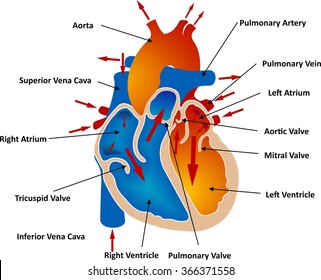
superior vena cava. inferior vena cava. right coronary artery. left coronary artery. posterior interventricular artery. anterior interventricular artery. circumflex artery. right marginal artery. small cardiac vein.
Heart Blood Flow Simple Anatomy Diagram Cardiac Circulation Pathway Steps Ezmed
December 1, 2020 - Aktivieren Sie JavaScript in den Browsereinstellungen oder wechseln Sie zu einem geeigneten Browser wie "Chrome" oder "Firefox", um via medici zu verwenden

Medical Definition Of Inferior Vena Cava
Recall that blood returning from the systemic circuit enters the right atrium via the superior and inferior venae cavae and the coronary sinus, which drains the blood supply of the heart muscle. These vessels will be described more fully later in this section.

Anatomy Of Major Abdominal Veins Inferior Vena Cava Segments Adapted Download Scientific Diagram
The inferior vena cava (IVC) is the largest vein in the body, draining blood from the abdomen, pelvis and lower extremities. This pictorial review summarises normal anatomy and embryological ...
The Inferior Vena Cava Anatomical Variants And Acquired Pathologies Insights Into Imaging Full Text
January 17, 2020 - Other articles where inferior vena cava is discussed: vena cava: Inferior vena cava.: The inferior vena cava is formed by the coming together of the two major veins from the legs, the common iliac veins, at the level of the fifth lumbar vertebra, just below the small of the back.

371 Inferior Vena Cava High Res Illustrations Getty Images
The inferior vena cava is formed by the joining of the common iliac veins which meet a little below the small of the back. The inferior vena cava travels along the spine, parallel to the aorta, and transports blood from the lower extremities of the body to the posterior region of the right atrium.

Diagram Showing The Rent In The Inferior Vena Cava And The Vascular Download Scientific Diagram
Inferior vena cava syndrome (IVCS) is a sequence of signs and symptoms that refers to obstruction or compression of the inferior vena cava (IVC). The pathophysiology of IVCS is similar to superior vena cava syndrome (SVCS) because of the presence of an underlying process that inhibits venous return to the right atrium. IVCS is not a primary diagnosis because it is often caused by other ...

371 Inferior Vena Cava High Res Illustrations Getty Images
The inferior vena cava then ascends to the right of the abdominal aorta along the vertebral column, receiving blood from numerous tributaries, and eventually passing through the caval foramen of the diaphragm. Notice that the veins draining the organs of gastrointestinal tract do not empty into the inferior vena cava.

Superior Vena Cava Inferior Vena Cava Coronary Sinus A Right Side Of The Heart B Left Side Of The Heart Study Com
15 Inferior Vena Cava Diagram. The inferior vena cava is the common convergence of venous drainage from all structures below the diaphragm. The inferior vena cava (ivc) drains venous blood from the lower trunk, abdomen, pelvis and lower limbs to the right atrium of the heart. In this image, you will find hepatic veins, inferior phrenic vein ...

Superior Vena Cava Wikipedia
Die Vena cava inferior (untere Hohlvene) ist die stärkste Vene des Körpers. Erfahre hier mehr zu ihrem Verlauf und dem Vena-cava-Kompressionssyndrom!
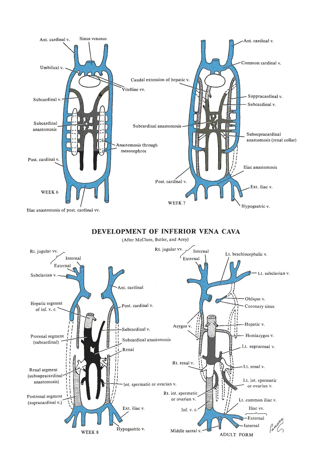
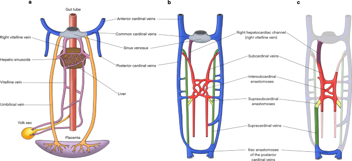
Chapter 126 Development Of The Venous System The Inferior Vena Cava Review Of Medical Embryology Book Lifemap Discovery
Overview of the inferior vena cava. The IVC is formed by the union of the right and left common iliac veins.It conveys systemic venous blood from the lower limbs and pelvis, the undersurface of the diaphragm and parts of the abdominal wall.The IVC does not drain blood from the gut.. Course of the IVC. The IVC begins in the abdomen at L5 and ends in the thorax at T8, where it enters the ...

Inferior Vena Cava And Its Tributaries Anatomy Tutorial Youtube
The ultrasound can assess fluid responsiveness as measure the maximal inferior vena cava diameter, inferior vena cava inspiratory collapse, and internal jugular aspect ratio. Amongst these three, the measurement of the maximal inferior vena cava diameter was found to be the best estimate of the central venous pressure with an inferior vena cava ...

Inferior Vena Cava Png Images Pngwing
Inferior Vena Cava. The IVC, a single right-sided vessel in 97% of individuals, returns blood from all structures below the diaphragm to the right atrium of the heart. It is formed by the confluence of the two common iliac veins at the level of the fifth lumbar vertebra just to the right of midline.

Urinary System Stock Vector Illustration Of Diagram 101625126
The inferior vena cava is a large heart vein that brings deoxygenated blood from parts of the lower body, including the legs, abdomen and pelvis, to the heart to be oxygenated. From the heart, this blood is then pumped back through the body to deliver oxygen to the body cells. Also known as the posterior vena cava, this large vein empties into ...

Inferior Vena Cava
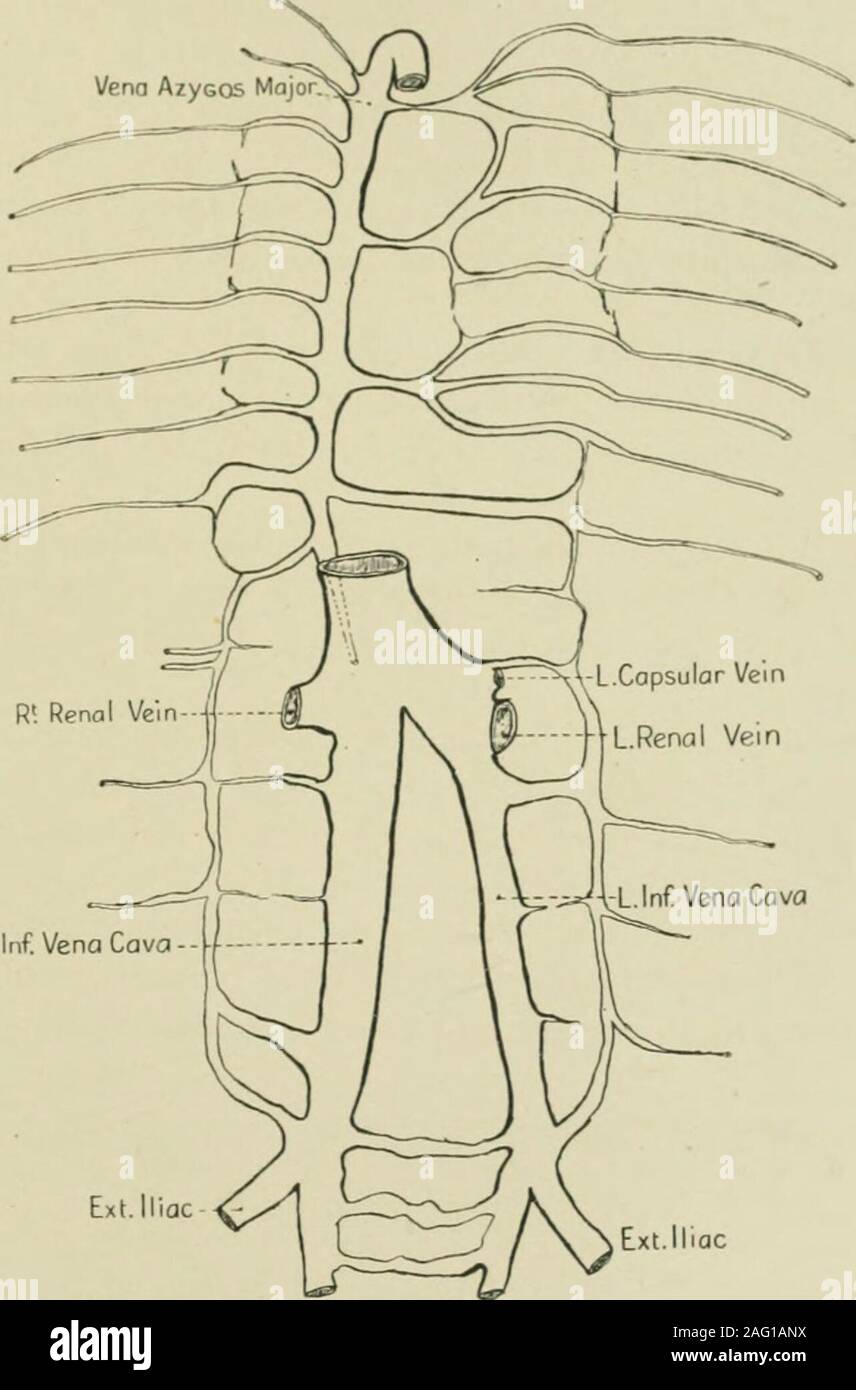
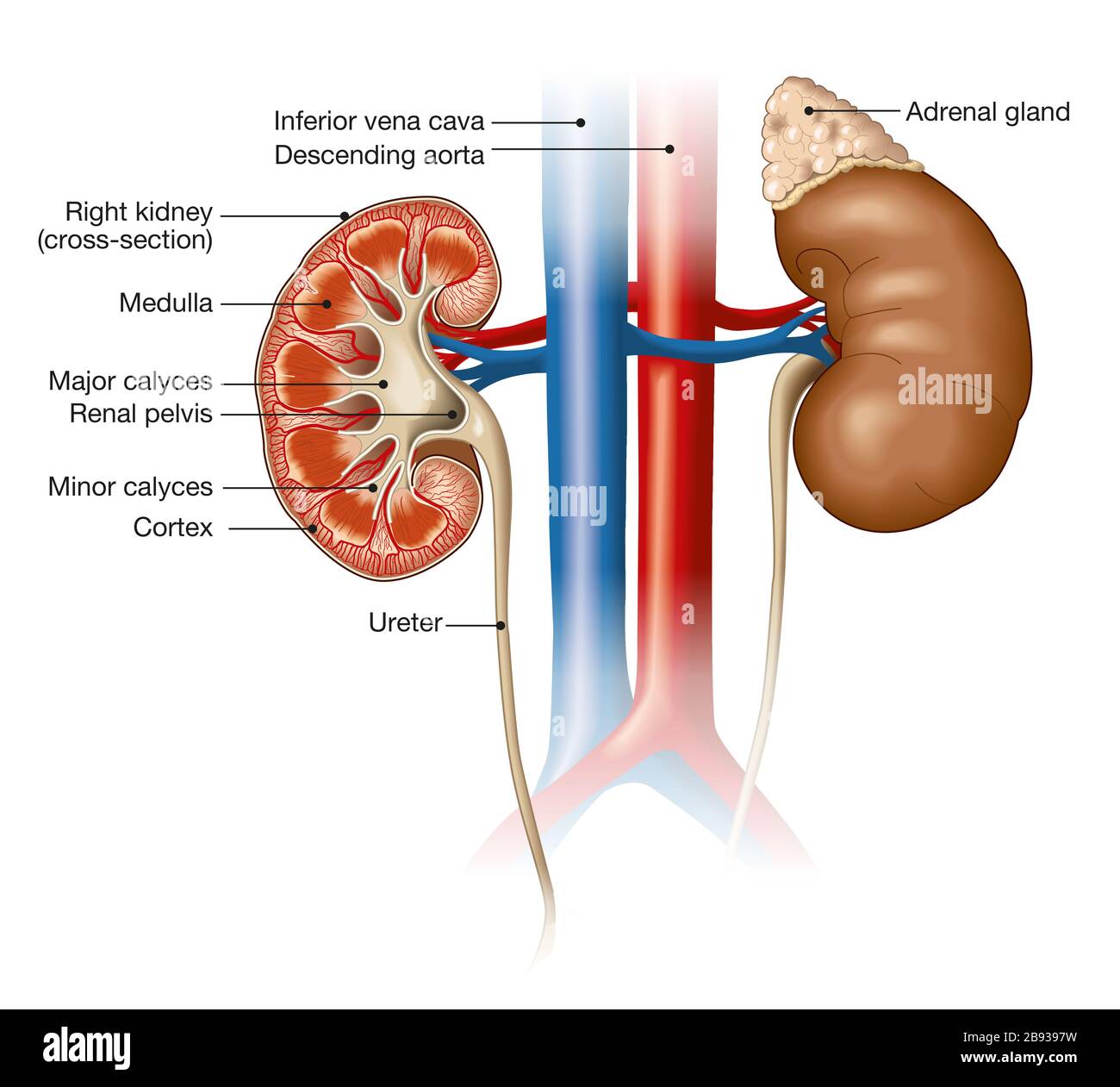
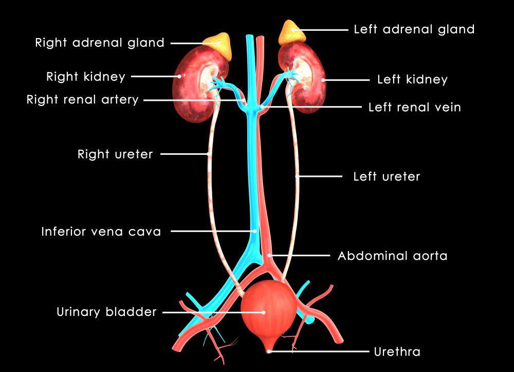
Inferior Vena Cava- Takes deoxygenated blood from the kidney back to the heart. Renal Artery- Takes oxygenated blood from the Abdominal Aorta to the kidneys. Renal Vain- Takes deoxygenated blood from the kidney to the Inferior Vena Cava. Kidneys- Filter waste and water together into urine. Ureter- tubes that take the urine to the bladder.
What Is The Superior Vena Cava With Pictures
August 31, 2020 - The inferior vena cava (IVC) is a large retroperitoneal vessel formed by the confluence of the right and left common iliac veins. Anatomically this usually occurs at the L5 vertebral level. The IVC lies along the right anterolateral aspect of the vertebral column and passes through the central ...

Diagram Word Bank Superior Vena Cava Inferior Vena Cava Right Ventricle Right Atrium Left Ventricle Aortic Arch Left Atrium Aorta Right Pulmonary Artery Ppt Download
Superior/Inferior Vena Cava. Now that we understand the blood flow to and from the heart, we can discuss the final structures. The first 2 structures are responsible for carrying deoxygenated blood from the body to the right side of the heart (right atrium). They are known as the superior vena cava and inferior vena cava.
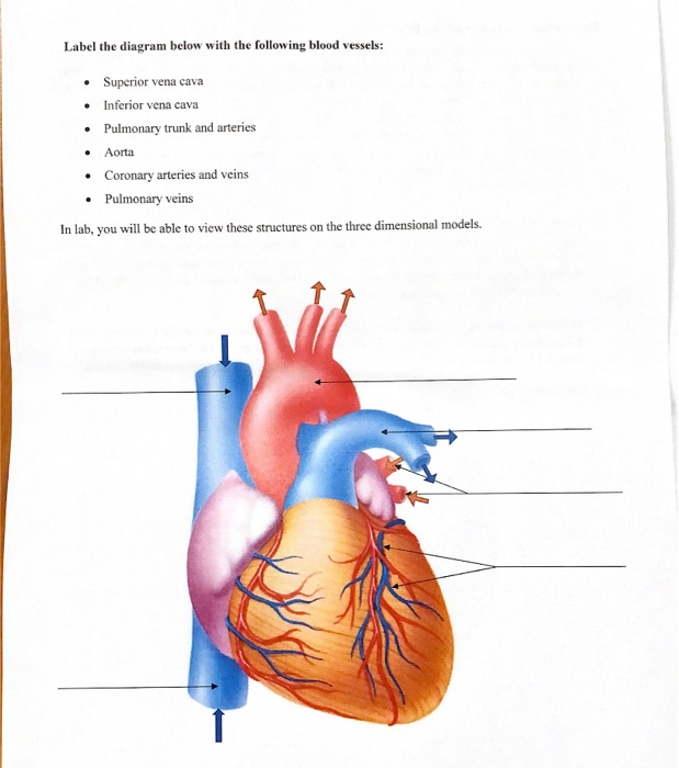
Solved Label The Diagram Below With The Following Blood Chegg Com
Inferior Vena Cava. The inferior vena cava is the largest vein in the human body. It collects blood from veins serving the tissues inferior to the heart and returns this blood to the right atrium of the heart. Although the vena cava is very large in diameter, its walls are incredibly thin due to the low pressure exerted by venous blood.

Inferior Vena Cava Diagram
tissues through the superior and inferior vena cava, pumps the blood into the right ventricle (RV) via the right atria ventricular orifice. RV then pumps the blood to the lungs for gas exchange, through the pulmonary trunk and arteries. Left atrium (LA) after receiving oxygenated blood from the lungs
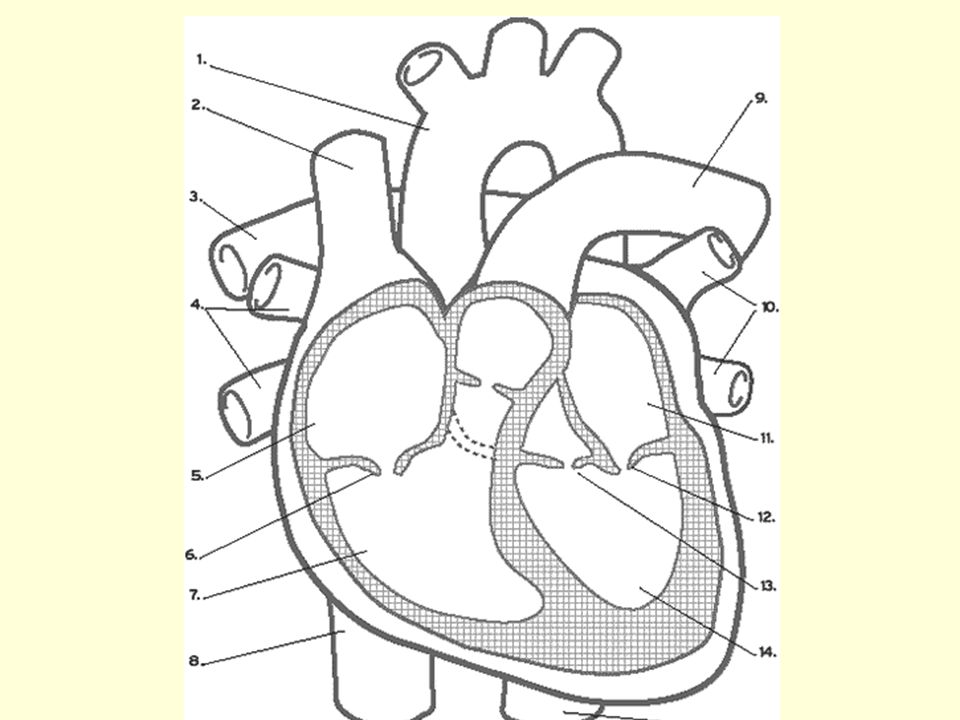
Diagram Word Bank Superior Vena Cava Inferior Vena Cava Right Ventricle Right Atrium Left Ventricle Aortic Arch Left Atrium Aorta Right Pulmonary Artery Ppt Download
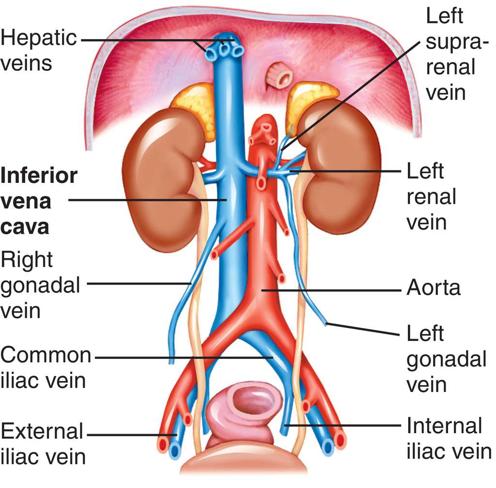
Inferior Vena Cava Diagram. In this image, you will find hepatic veins, inferior phrenic vein, portal vein, left renal vein, left suprarenal vein, left gonadal vein, right gonadal vein, right renal vein in it. You may also find right suprarenal vein, aorta, left common iliac vein, right common iliac vein, left external iliac vein, median sacral ...
Inferior Vena Cava High Resolution Stock Photography And Images Alamy
The inferior vena cava (IVC) (plural: inferior venae cavae) drains venous blood from the lower trunk, abdomen, pelvis and lower limbs to the right atrium of ...Missing: diagram | Must include: diagram
What Is The Superior Vena Cava With Pictures
Locate the inferior vena cava and superior vena cava. 7. Use the scalpel to open the heart chambers: make an incision along the coronal plane, from the superior portion of the left ventricle to the superior portion of the right ventricle. Inferior Vena Cava Superior Vena Cava Posterior View Vena Cava = Singular Vena Cavae = Plural NOTICE the ...

Inferior Vena Cava Anatomy Britannica
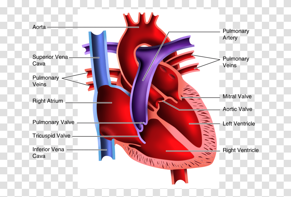
blood flow enters right atrium from superior/inferior vena cava and coronary sinus through the tricuspid valve into the right ventricle and then through the pulmonic valve to the pulmonary artery into the lungs and collected by. pulmonary veins. blood flow THROUGH the heart starts by.
1
Vena renalis dextra et sinistra: Die linke Nierenvene nimmt i.d.R. die Vena suprarenalis sinistra, die linken Venae testiculares bzw. Venae ovaricae sowie die linke Vena phrenica inferior auf. ... Vena testicularis bzw. Vena ovarica dextra ... Zwischen der Vena cava inferior und der Vena cava ...

Inferior Vena Cava Png Pngegg
Step 1 involves the superior vena cava (SVC) and inferior vena cava (IVC). They are the main blood vessels that carry the deoxygenated venous blood from the rest of the body to the right side of the heart, specifically the right atrium. The superior vena cava is located superiorly, and it carries the deoxygenated venous blood from the upper ...
Full Size Picture Inferior Vena Cava Jpg
August 5, 2016 - Die obere und untere Hohlvene (Vena cava superior, Vena cava inferior) sind die zwei größten Venen im menschlichen Körper. Sie sammeln sauerstoffarmes Blut aus der Körperperipherie und leiten es zurück zum Herzen, genauer gesagt in den rechten Herzvorhof.
Vena Cava Images Stock Photos Vectors Shutterstock
6 Inferior Vena Cava. The great vessels of the heart function to carry blood to and from the heart as it pumps, located largely within the middle mediastinum. In this article we will consider the structure and anatomical relationships of the aorta, pulmonary arteries and veins, and the superior and inferior vena cavae.

File Diagram Of The Human Heart Cropped It Png Wikimedia Commons

Heart Valve Atrium Anatomy Diagram Heart Purple Text Hand Png Pngwing

Biophysical Science Biophysics Superior Aorta Vena Cava Pulmonary Artery Right Auricle Inferior Vena Cava Pulmonary Vein Left Auricle Incomplete Septum A B Figure 3 Diagrams Offish And Reptile Hearts A Fish
Human Heart Clipart Blood Flow Human Heart Pictures For Students Plot Diagram Dynamite Bomb Transparent Png Pngset Com
Hcs 1080 The Cardiovascular System Diagram Of The

Instant Anatomy Abdomen Vessels Veins Inferior Vena Cava
Inferior Vena Cava Anatomy And Function Kenhub
Inferior Vena Cava Anatomy And Function Kenhub
0 Response to "37 inferior vena cava diagram"
Post a Comment