39 hip flexor anatomy diagram
Diagram Of Hip Muscles And Ligaments Move your left leg back up until the top of your thigh rests on the ground. Using your hands, carefully push up till your spine is straight. To deepen the position, place your lower arms on the ground and lean forward from your hips. If the hip flexors are tight, the resulting anterior pelvic tilt and lumbar hyperextension will cause problems in many yoga poses, including standing poses like Virabhadrasana II (Warrior Pose II) and Trikonasana (Triangle Pose), in which the primary leg action is opening to the sides instead of flexing forward or extending back.
Hip flexor strain occurs when you use your hip flexor muscles and tendons too much. As a result, the muscles and tendons become inflamed, sore, and painful. Some people are more likely than others ...
Hip flexor anatomy diagram
3D Muscle Premium 2 by Visible Bodyhttp://goo.gl/9rcmed (Amazon Link)Ryan's Deadlift Nerd Newsletterhttp://deadliftnerd.com/deadlift-nerd-newsletter/Follow m... Hip joint (Articulatio coxae) The hip joint is a ball and socket type of synovial joint that connects the pelvic girdle to the lower limb. In this joint, the head of the femur articulates with the acetabulum of the pelvic (hip) bone.. The hip joint is a multiaxial joint and permits a wide range of motion; flexion, extension, abduction, adduction, external rotation, internal rotation and ... Hip Diagram Slide your left leg back until the top of your thigh rests on the ground. Utilizing your hands, gently push up until your spine is straight. To deepen the posture, place your lower arms on the ground and lean forward from your hips. Depending upon your versatility, you may be able to rest your forehead on the ground.
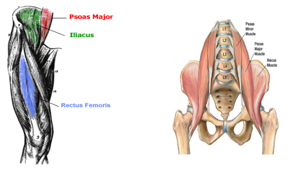
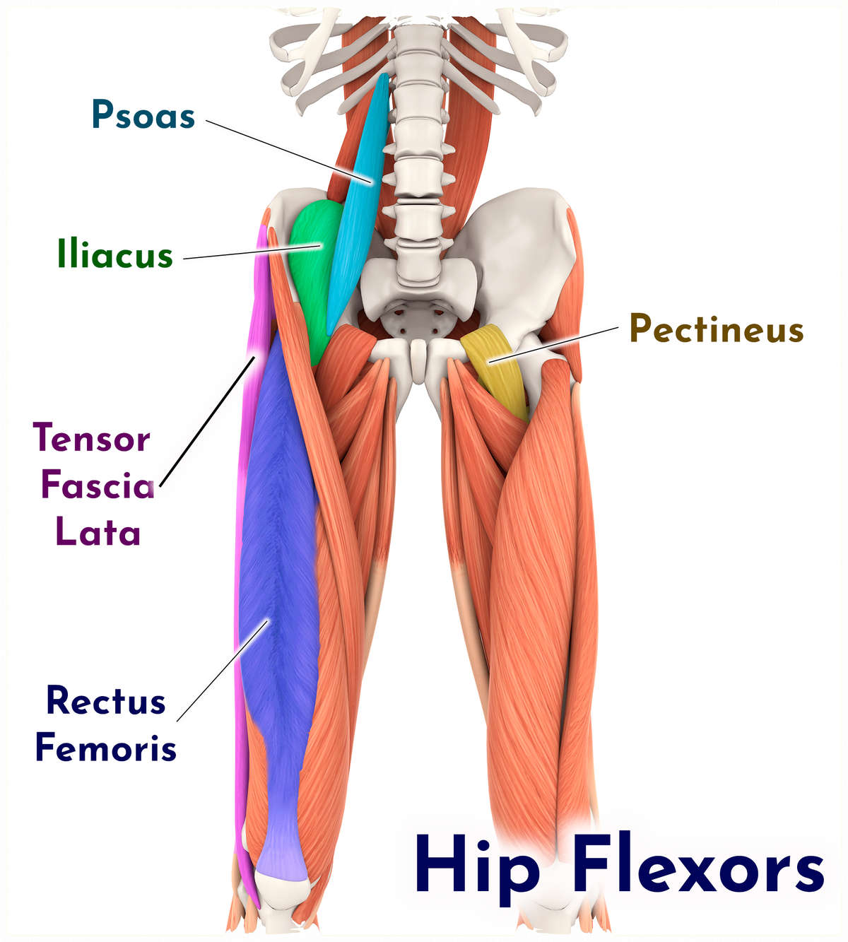
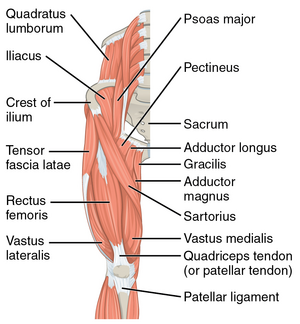
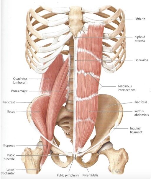
Hip flexor anatomy diagram. rotation at the hip and 90° flexion at the knee, the muscle becomes prominent and is easily palpable. The rectus femoris combines movements of flexion at the hip and extension at the knee. Its origin is at the anterior . inferior iliac spine, a groove above the acetabulum and the fibrous capsule of the hip joint and inserts into the common Beginner hip flexor muscle anatomy. If you're just starting your anatomy journey, work on remembering the names of all 11 hip flexor muscles. Use acronyms to help you. Here are the letters to work with: AAA I GG PP R S T. Scroll down to see the muscle names that go with these letters. Hip Anatomy Diagram: From Bones To Joints. ... General Hip Anatomy. The hip is a ball-and-socket joint, similar to the joint in the shoulder. Part of the reason for the hip's stability is that there is a very deep socket, called the acetabulum, in the hip joint. A strong capsule joint supported by ligaments and muscles also provides extra ... Hip Flexors: Psoas, and Iliacus. This drawing illustrates the pathway of your psoas. Ignore the fact that "major" and "minor" are both listed. These muscles contract strongly in postures like navasana and bakasana. They stretch in all of your backbends and lunges. Hip Flexors: Your Rectus Femoris. The rectus femoris is a "double-duty ...
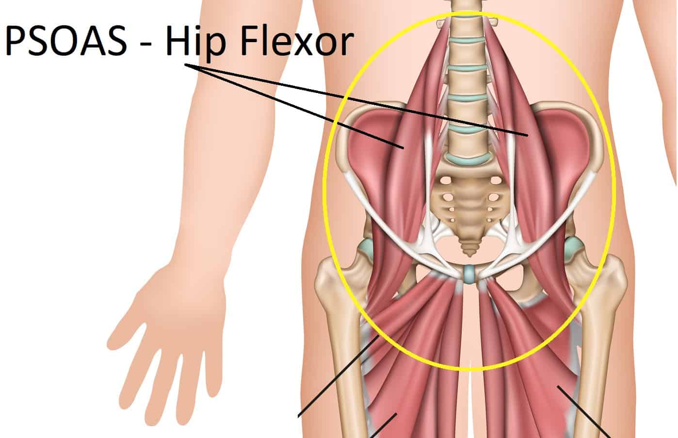
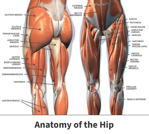
The hip flexor has both major and minor functions, it is able to fulfill different roles because it is composed of several muscles, the largest ones are discussed below. The primary goal of the hip flexor is to facilitate flexion of the hip joint. In normal terms, this means that the hip flexor is used anytime the knee is lifted up, a step is ... The iliopsoas muscle is a major mover of your hip joint. It's formed by the joining of three muscles: the iliacus muscle, the psoas major muscle, and the psoas minor muscle. These muscles work together to flex your hip and to stabilize your hip and lower back during activities such as walking, running, and rising from a chair. The hip muscles encompass many muscles of the hip and thigh whose main function is to act on the thigh at the hip joint and stabilize the pelvis.Without them, walking would be impossible. They can be divided into three main groups: Iliopsoas group; Gluteal muscles; Hip adductors; This article will introduce the muscles in each group and touch on their origin, insertion, function, and innervation. The hip joint is one of the most flexible joints in the entire human body. The many muscles of the hip provide movement, strength, and stability to the hip joint and the bones of the hip and thigh. These muscles can be grouped based upon their location and function. The four groups are the anterior group, the posterior group, adductor group ...
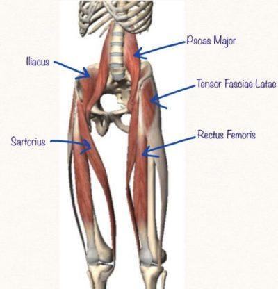
Hip flexor pain can be either acute, arising from trauma, or chronic, becoming worse over time. Common symptoms of hip flexor tendinosis include: Pain at the front of the hip or in the groin. Pain while walking or climbing stairs. Pain when lifting the knees toward the chest. Altered gait or limping due to hip or groin pain Hip Flexor Muscles Anatomy. The hip flexor is not one singular muscle but a group of muscles, including the psoas major, iliacus, rectus femoris, pectineus, and sartorius. (The rectus femoris is also considered a quadricep muscle.) These muscles attach to various points on the spine, pelvis, and femur (thigh bone). Live. •. This week we are returning to our discussion of 'the basic anatomy' series and we are taking a look at the hip flexors, but specifically the psoas and iliacus muscles. The hip flexors are all the muscles that flex the hip joint. Surprise! There are 4: sartorius, rectus femoris, iliacus, and psoas. Commonly the psoas and iliacus are ... The hip and leg are sore and should not be massaged excessively, but the arm muscles are fine. By activating the extensors in the right shoulder and arm, the left hip extensor muscles can be facilitated. By activating the flexors of the left arm, the flexors of the left hip are inhibited. This process may restore balance in the gait pattern.
Hip Flexor Muscle
Hip Abduction Exercises Anatomy Benefits Effectiveness from i0.wp.com It attaches inferiorly (underneath/below) to the long thick strip of fascia, known as. There are various hip flexor muscles that all work to. See more ideas about anatomy, massage therapy, hip anatomy. The thigh bone or femur and the pelvis join to form the hip joint.
Why The Psoas Is Significant
Hip flexor stretch Standing in a wide walking position, a person should put both hands on a firm support in front of them. Lunge forward and bend the front knee. They should push their hips forward...

Why Hip Flexors Are Tight and Why Your Hips Pop | Sparta Science
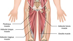
Adductor muscles on the inside of your thigh. Iliopsoas muscle, a hip flexor muscle that attaches to the upper thigh bone. Rectus femoris muscle, one of the quadriceps muscles on the front of your...
5 Hip Flexor Stretches!
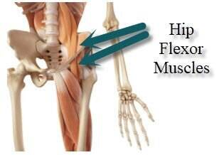
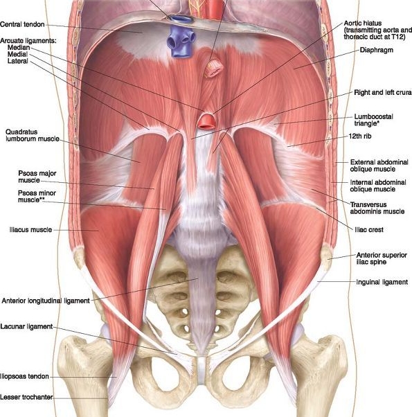
The muscles that sit at the front of the hip are called the hip flexors (Figure 2.2) and act to lift your knee towards your chest (flexion). The main hip flexor, the iliopsoas muscle is made up of two parts - the psoas muscle that starts at the lumbar spine, and the iliacus that starts from the inside of the pelvis.

How To Unlock Tight Hip Flexors - EMPOWER YOUR WELLNESS
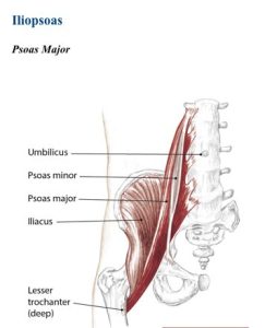
Anatomy of the Hip Flexor Muscles - Iliacus and the Psoas Major, Iliopsoas, Rectus Femoris Hip Flexor Muscle Anatomy The Iliopsoas actually consists of two muscles: the Iliacus and the Psoas Major. Together, they are known as the Iliopsoas. Anatomy Chart courtesy of FCIT The Iliacus originates on the pelvic crest and attaches on the femur.

Psoas major Part I: hip flexor or lumbar stabilizer?
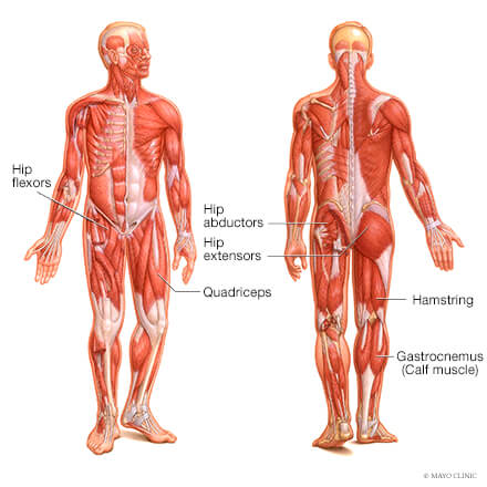
The hip flexors are several muscles that bring your legs and trunk together in a flexion movement. They allow you to move your leg or knee up towards your torso, as well as to bend your torso forward at the hip. You can strain or tear your hip flexor muscles through sudden movements or falls. 1.

Basic Anatomy of Stretching the Hip Flexors - Movement Fix
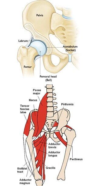
The hip joint is a ball-and-socket type jointand is formed where the thigh bone (femur) meets the pelvis. The femur has a ball-shaped head on its end that fits into a socket formed in the pelvis, called the acetabulum. Large ligaments, tendons, and muscles around the hip joint hold the bones (ball and socket) in place and keep it from dislocating.

How To Unlock Tight Hip Flexors - EMPOWER YOUR WELLNESS
Motions Available. The hip joint connects the lower extremities with the axial skeleton. The hip joint allows for movement in three major axes, all of which are perpendicular to one another. The location of the center of the entire axis is at the femoral head. The transverse axis permits flexion and extension movement.
Fix Hips to Ease Back Pain — Evolve Fitness Chicago
Hip Diagram Slide your left leg back until the top of your thigh rests on the ground. Utilizing your hands, gently push up until your spine is straight. To deepen the posture, place your lower arms on the ground and lean forward from your hips. Depending upon your versatility, you may be able to rest your forehead on the ground.

How To Get Immediate Psoas Pain Relief - Coach Sofia Fitness
Hip joint (Articulatio coxae) The hip joint is a ball and socket type of synovial joint that connects the pelvic girdle to the lower limb. In this joint, the head of the femur articulates with the acetabulum of the pelvic (hip) bone.. The hip joint is a multiaxial joint and permits a wide range of motion; flexion, extension, abduction, adduction, external rotation, internal rotation and ...

Understand Hip Anatomy Muscles for Yoga | Jason Crandell Yoga
3D Muscle Premium 2 by Visible Bodyhttp://goo.gl/9rcmed (Amazon Link)Ryan's Deadlift Nerd Newsletterhttp://deadliftnerd.com/deadlift-nerd-newsletter/Follow m...

The Hip Flexors – Wellbeing Physiotherapy, Massage and ...

Deep Dive into the Anatomy of the Hip Flexor Muscles
Hip Flexors Advanced Level – EasyFlexibility

Hip Pain Explained - including structures & anatomy of the ...
Hip Muscles - Origin, Insertion, Action and Exercises ...

Muscles of the Hip - Anatomy Pictures and Information

Hip Pain Explained - including structures & anatomy of the ...
Understanding the Airway Part 3: The Diaphragm — Hands On ...
The New Psoas Stretch - Try this video for a new take on a ...

Joburg Post

Deep Dive into the Anatomy of the Hip Flexor Muscles
Hip Flexors - Physiopedia

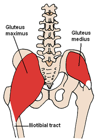
Anatomy Lesson: The Hips and Glutes - Goodwin House
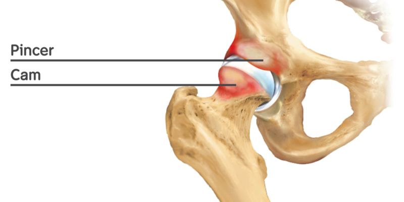
Hip Impingement Vs Hip Flexor Strain - Rush Chiropractic Center
Muscles of the Hip Joint
Hip Pain and Tightness? inTune Chiropractic is Here with Some ...
Muscles of the hip - Wikipedia
💪ðŸ¼ðŸ’ªðŸ¿PT CORNER: Hip Flexor Pain 💪ðŸ¿ðŸ’ªðŸ¼ — Fierce45®

Hip Flexor Strain – Considerations and Understanding
What is a Hip Flexor? - Plano Orthopedic & Sports Medicine Center
Hip Flexor Strains | Florida Orthopaedic Institute
Hip Strains - OrthoInfo - AAOS

Psoas Hip-Flexors - Home | Facebook

86 Hip anatomy ideas | anatomy, massage therapy, muscle anatomy

Deep Dive into the Anatomy of the Hip Flexor Muscles

Hip muscles, lateral view. | Hip muscles anatomy, Hip muscles ...
![Hip Flexor Ability Anatomy Review [Psoas, Rectus Femoris, Quads]](https://i.ytimg.com/vi/UJyyVf9PDg4/hqdefault.jpg)
Hip Flexor Ability Anatomy Review [Psoas, Rectus Femoris, Quads]
Muscle strains (IT band, groin, hip flexor) - Mayo Clinic ...
0 Response to "39 hip flexor anatomy diagram"
Post a Comment