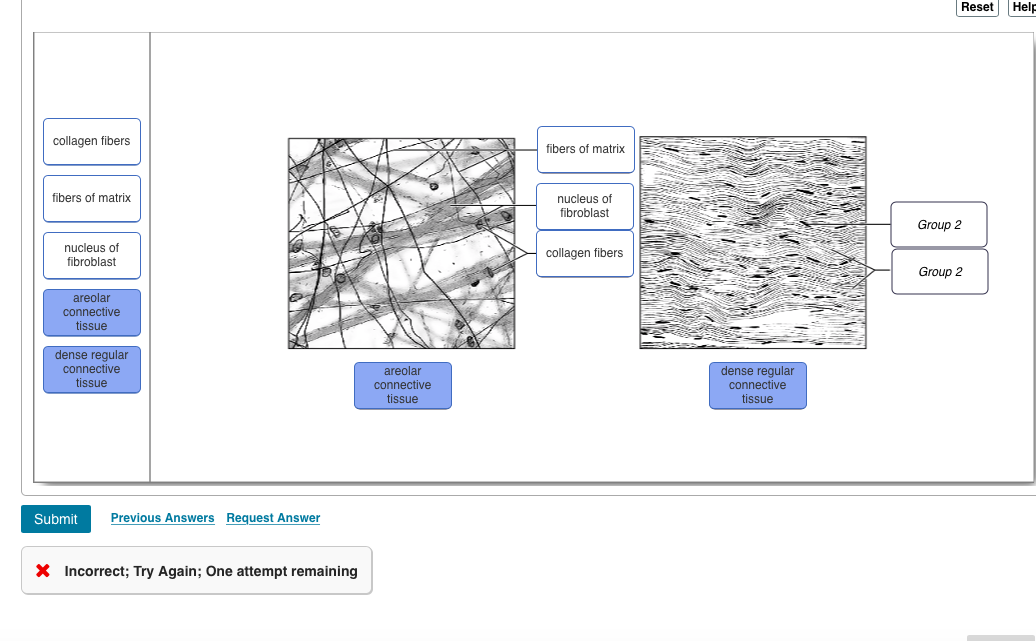
37 drag the labels onto the diagram to identify the types of connective tissue proper.
Part A Drag the labels onto the diagram to identify the structures of the neuron. ... Correct Chapter 4 Multiple-Choice Question 31 Part A Which of the following is a type of connective tissue? ANSWER: cilia apical surface microvilli basement membrane stratified squamous. stratified cuboidal. simple cuboidal. transitional. simple squamous. Drag the appropriate labels to their respective targets. back 3. ... Connective tissue sacs lined with synovial membranes that act as cushions in places where friction develops are called _____. ... Identify the saddle joint of the skeleton. back 29. Carpometacarpal joint of the thumb.
Drag the labels onto the diagram to identify the types of supporting connective tissues. look at pic A marked loss in strength and elasticity of connective tissue characterizes Marfan's syndrome.
Drag the labels onto the diagram to identify the types of connective tissue proper.
Identify the connective tissue proper cellular component labeled "F". ... Drag the labels onto the diagram to identify the muscle types based on fascicle ... Connective tissue Basement membrane Nucleus IDIO Lumen of duct S tified cuboidal cells Submit Request Answer 1301051aafd2 787. This problem has been solved! See ... Drag the labels onto the diagram to identify the types of epithelia. The polarity of epithelial cells identify the structures in epithelial cells. Drag the labels onto the diagram to identify the stages in which the lagging strand is synthesized. The layers do not contain any blood vessels and one surface of the cells lines the cavity of the organ.
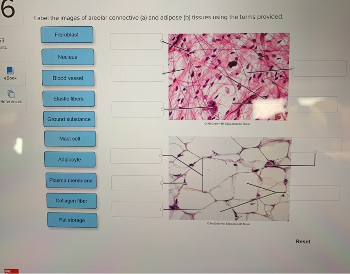
Drag the labels onto the diagram to identify the types of connective tissue proper.. Question: Drag the labels onto the diagram to identify the components of the integumentary system. Reset Stre Cutaneous embra Har shaft Pore swear gland duct Talle COCOLISCIA Seondecus gland. rectomp muscle Hair follicle cam QUISA Swearglana du Sweat gland Nerve fibers Submit Previous Answers Request Answer . Connective Tissue Disorder. Connective tissue disorders may be due to mutation of genes or by inherited faulty genes. The two genetic disorders of connective tissue are epidermolysis bullosa (EB) and Marfan syndrome. Marfan syndrome is due to defective genes producing a protein fibrillin-1. The disease is characterised by a very thin and long body. Drag the labels onto the diagram to identify the cell types and matrix components of areolar connective tissue, a model connective tissue. Of the two major cell types found in nervous tissue, __________ cells are highly specialized to generate and conduct electrical signals. Part A Drag the labels onto the diagram to identify the types of supporting connective tissues. ANSWER: Correct Chapter 4 Book-specific Clinical Case Activity All her life Anne Marie has felt "delicate." She is a horrible athlete. She once dislocated both shoulders attempting to play high school volleyball. And she has an unstable feeling, as if her kneecaps will dislocate when she tries ...
Image: Drag the labels onto the diagram to identify the types of connective tissue proper. The structure of bone tissue suits the function. Drag the labels onto the diagram to identify structural features associated with skeletal muscle. Study questions on anatomy review. Drag the appropriate labels to their respective targets. Drag the labels onto the diagram to identify the blood types that correspond to specific blood typing test ... connective tissue: A type of tissue found in animals whose main function is to bind, support, and anchor the body. Connective tissue (CT) is a one of the four main classes of tissues. Although it is the most abundant and widely distributed of the primary tissues, the amount of connective tissue in a particular organ varies. Correct Exercise 6 Review Sheet Art labeling Activity 1 Identify the tissues from INTEGBI 1 at University of California, Berkeley
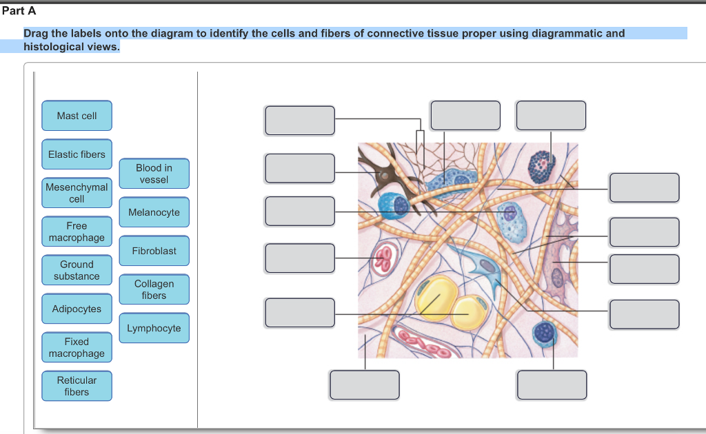
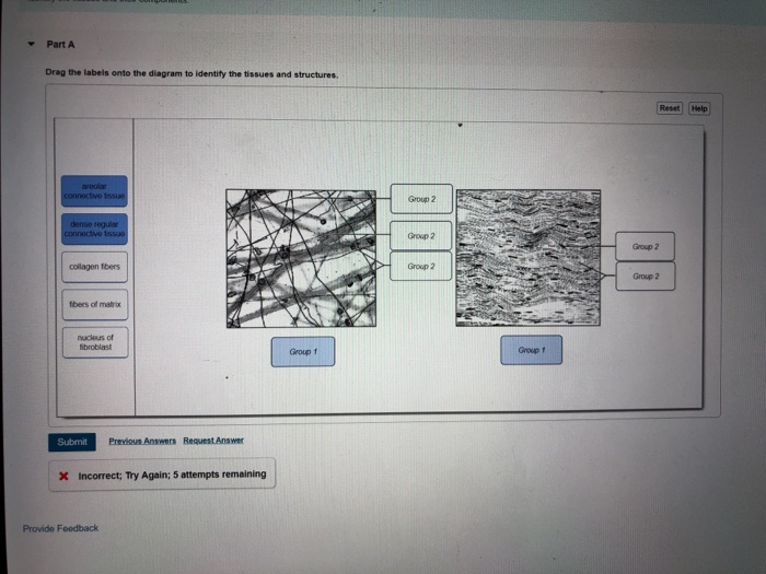
Drag the labels onto the diagram to identify the cells and fibers of connective tissue proper using diagrammatic and histological views. This problem has been ... Anatomy and Physiology questions and answers. Drag the labels onto the diagram to identify structures of the urinary bladder. Re Basal lamina Connective tissue Stretched bladder Epithelium (stretched) Relaxed bladder Epithelium (relaxed) Question: Drag the labels onto the diagram to identify structures of the urinary bladder. Elastic connective tissue is a modified dense connective tissue that contains numerous elastic fibers in addition to collagen fibers, which allows the tissue to return to its original length after stretching Figure 4.10). The lungs and arteries have a layer of elastic connective tissue that allows the stretch and recoil of these organs. Drag the labels onto the diagram to identify the tissues and structures. Reset Help central cand matrix Group 2 lacuna ... Cartilage and bone are two specialized types of connective tissues. ... This tissue is rigid but provides flexible support to specific regions. It places in areas where there is constant and direct compression.
Answered A Session Masteringaandp Com E Bartleby
Drag the labels onto the diagram to identify the types of connective tissue proper. look at pic When a molecule is _______ it loses electrons, and when a molecule is ______ it gains electrons.

Types Of Tissues Anatomy And Physiology I
Question: Part A Drag the labels onto the diagram to identify the cells and fibers of connective tissue proper using diagrammatic and histological views.
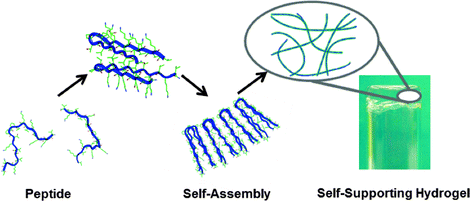
Peptide Based And Polypeptide Based Hydrogels For Drug Delivery And Tissue Engineering Springerlink
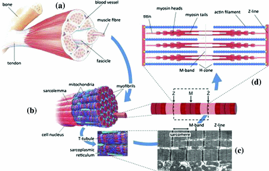
Each skeletal muscle has three layers of connective tissue that enclose it, provide structure to the muscle, and compartmentalize the muscle fibers within the muscle (Figure 10.2.1). Each muscle is wrapped in a sheath of dense, irregular connective tissue called the epimysium , which allows a muscle to contract and move powerfully while ...

Pdf Multiparametric Analyses Of Hepatocellular Carcinoma Somatic Mouse Models And Their Associated Tumor Microenvironment
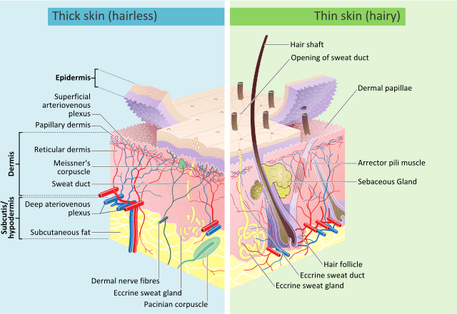
Drag the appropriate labels to their respective targets. 2. ... The dense fibrous connective tissue portion of the skin is located in the reticular region of the dermis. ... glands are categorized as two distinct types. Which of the following are the two types of sweat glands? eccrine and apocrine. 56. Which layer(s) of the skin is(are) damaged ...

Nanoparticles Influence In Skin Penetration Of Drugs In Vitro And In Vivo Characterization Sciencedirect
Identify the tissue type and a location where it is found. Loose Areolar Connective Tissue •Papillary layer of dermis • Hypodermis •Around organs • Basement membrane of mucous membranes •Surrounding blood vessels Blood Vessel. Identify the tissue type and its function.

Ercise 6 Review Sheet Art Labeling Activity 1 Drag The Labels Onto The Diagram To Identify The Homeworklib
The word tissue derives from the Old French word meaning "to weave," reflecting the fact that the different tissues are woven together to form the "fabric" of the human body. The four basic types of tissue are epithelial tissue, connective tissue, muscle tissue, and nervous tissue. If a single, broad functional term were assigned to ...
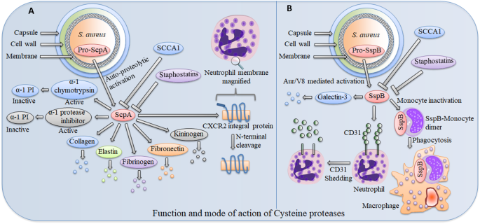
Interaction Of Host And Staphylococcus Aureus Protease System Regulates Virulence And Pathogenicity Springerlink
Part A Drag the labels onto the diagram to identify the cells and fibers of ... Answer There types of tissues included in connective tissue proper are ...
Buffaloschools Org
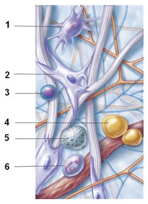
(c) Connective tissue proper: loose connective tissue, reticular Description: Network of reticular fibers in a typical loose ground substance; reticular cells lie on the network. Function: Fibers form a soft internal skeleton (stroma) that supports other cell types including white blood cells, mast cells, and macrophages.
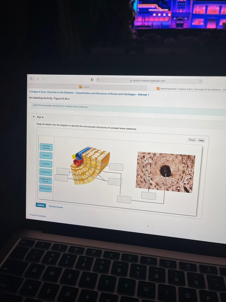
35 Drag The Correct Label To The Appropriate Location To Identify The Parts Of An Osteon Labels Design Ideas 2020
Correct Reading Quiz - Chapter 4 Question 1 Part A Which of these is not one of the four recognized tissue types? ANSWER: Correct Chapter 4 Question 85 Part A The study of tissues is called _____. ANSWER: Correct Art Activity: The Polarity of Epithelial Cells Identify the structures in epithelial cells. Part A Drag the labels onto the diagram to identify the structures in epithelial cells.
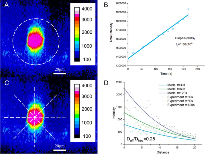
Modulation Of Solute Diffusivity In Brain Tissue As A Novel Mechanism Of Transcranial Direct Current Stimulation Tdcs Scientific Reports
Areolar tissue, found in the hypodermis of the skin and below the epithelial layers of the digestive, respiratory, and urinary tracts, is a loose connective tissue proper, as is adipose tissue, also known as fat. Table 5.1 lists some of the subcategories of connective tissue proper, along with some of their characteristics and properties.
Solved Drag The Labels Onto The Diagram To Identify The Chegg Com
Drag the labels onto the diagram to identify the cells and fibers of connective tissue proper using diagrammatic and histological views. Image: Reticular Fibers ...
Of Limbs Wings And Fins Springerlink
These tissues include the skeletal muscle fibers blood vessels nerve fibers and connective tissue. Drag the labels onto the diagram to identify structural features associated with skeletal muscle. Drag the labels onto the diagram to identify the blood types that correspond to specific blood typing test results.
Integumentary System Definition Organs Functions Diseases
Drag the labels onto the diagram to identify the types of connective tissue proper. (week three assignment one) #7. Drag the labels onto the four types of tissue membranes. ... Drag the labels onto the diagram to identify the effects of isotonic, hypotonic, and hypertonic solutions on red blood cells.

Chapter 47 Sensory Perception Bio 140 Human Biology I Textbook Libguides At Hostos Community College Library
Connective tissue is a term used to describe the tissue of mesodermal origin that that forms a matrix beneath the epithelial layer and is a connecting or supporting framework for most of the organs of the body. This lab will focus on the so-called connective tissue proper and cartilage; the next lab will focus on bone.

Lymphatic Vessel Network Structure And Physiology Abstract Europe Pmc
Chapter 4 Notes Epithelial and Connective Tissue. Histology: the study of tissues. 1. Definition. a tissue is a group of similar cells with intercellular material that together perform a specific function.

The Biofilm Adhesion Protein Aap From Staphylococcus Epidermidis Forms Zinc Dependent Amyloid Fibers Journal Of Biological Chemistry
For page 1, the answers are:- Aerolar tissue Adi …. View the full answer. Transcribed image text: -Aibrocytes Reticular fbers bundles Part A Drag the labels onto the diagram to identify the types of supporting connective tissues Chondrocytes in lacunae Chondrocytes in lacunae Matrix Elastic fibers Böne Osteocytes in lacuna Chondrocytes in ...
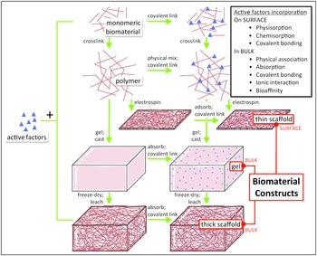
Enabling Technologies For Regenerative Pharmacology Section Ii Regenerative Pharmacology
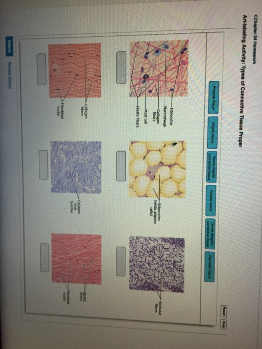
Part A Drag the labels onto the diagram to identify the types of epithelia. ANSWER: Correct Art-labeling Activity: Types of Connective Tissue Proper Learning Goal: To learn the types of connective tissue proper. Label the types of connective tissue proper.
Drag The Labels Onto The Diagram To Identify The Parts Of The Cell Wiring Site Resource
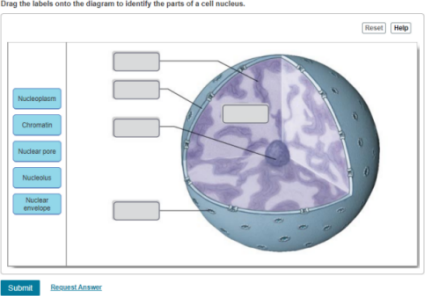
Drag the correct description under each cell structure to identify the role it plays in the cell. Part a animal cell structure drag the labels onto the diagram to identify the structures of an animal cell. Drag the labels onto the diagram to identify the parts of the cell. Labels can be used once more than once or not at all.

2017 Pearson Education Inc Ppt Download
Drag the labels onto the diagram to identify the types of connective tissue proper. areolar tissue, adipose tissue, reticular tissue, dense regular connective tissue, dense irregular connective tissue, elastic tissue. Myosatellite cells are found in association with _____ muscle.
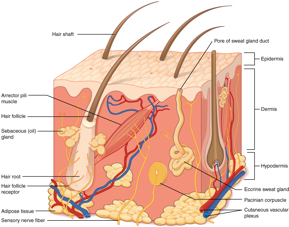
Layers Of The Skin Anatomy And Physiology
Drag the labels onto the diagram to identify the types of epithelia. The polarity of epithelial cells identify the structures in epithelial cells. Drag the labels onto the diagram to identify the stages in which the lagging strand is synthesized. The layers do not contain any blood vessels and one surface of the cells lines the cavity of the organ.
Mdpi Com
Connective tissue Basement membrane Nucleus IDIO Lumen of duct S tified cuboidal cells Submit Request Answer 1301051aafd2 787. This problem has been solved! See ...

Drag The Labels Onto The Diagram To Identify The Cells And Fibers Of Connective Course Hero
Identify the connective tissue proper cellular component labeled "F". ... Drag the labels onto the diagram to identify the muscle types based on fascicle ...
Lamission Edu

Structure And Function Of Blood Vessels Anatomy And Physiology

Ch 5 7 Lab A P Mastering Flashcards Quizlet
Solved Drag The Labels Onto The Diagram To Identify The Chegg Com

Bone Physiology As Inspiration For Tissue Regenerative Therapies Abstract Europe Pmc

Bone Physiology As Inspiration For Tissue Regenerative Therapies Abstract Europe Pmc
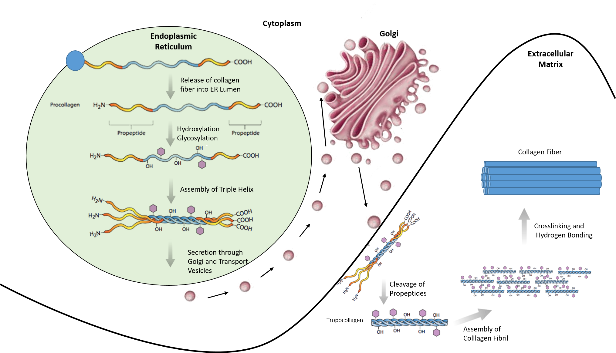
Chapter 2 Protein Structure Chemistry

Abstracts From Attd 20147th International Conference On Advancedtechnologies Treatments For Diabetesvienna Austria February 5 8 2014 Diabetes Technology Therapeutics
Solved Drag The Labels Onto The Diagram To Identify The Chegg Com
1
Solved Aibrocytes Reticular Fbers Bundles Part A Drag The Chegg Com

Intimal And Medial Contributions To The Hydraulic Resistance Of The Arterial Wall At Different Pressures A Combined Computational And Experimental Study Journal Of The Royal Society Interface

Pdf Multiparametric Analyses Of Hepatocellular Carcinoma Somatic Mouse Models And Their Associated Tumor Microenvironment
Solved Classify The Body Location Structure With The Correct Chegg Com
Answered A Session Masteringaandp Com E Bartleby
0 Response to "37 drag the labels onto the diagram to identify the types of connective tissue proper."
Post a Comment