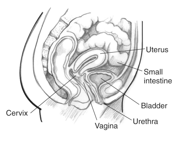
41 diagram of uterus and bladder
A Gallery of High-Resolution, Ultrasound, Color Doppler & 3D Images... Congenital anomalies of uterus. Uterus didelphys or Didelphic uterus. These ultrasound images are diagnostic of uterus didelphys or didelphic uterus. This is an extreme example of complete failure of fusion of the 2 halves of the uterus (Mullerian ducts) during the fetal stage. The Urogenital System | Veterian Key | Urinary Bladder FIGURE 9-8 Dorsolateral contrast radiograph of the bladder and ureters. The abdominal part of the FIGURE 9-32 Diagram of venous pathways in the bulbus glandis connecting the deep vein of the The uterus contained four embryos in the left horn and three in the right, indicating that at least two...
Anatomy and Physiology of Animals/Reproductive System - Wikibooks... Diagram 13.3 - Diagram summarizing the functions of the male reproductive organs. The two sperm ducts join the urethra just below the bladder, which passes through the penis and transports both sperm These maintain the lining of the uterus and prepare the mammary glands for milk secretion.
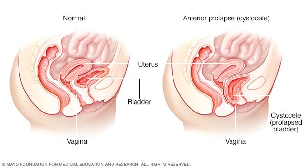
Diagram of uterus and bladder
Urinary Bladder - Earth's Lab Urinary Bladder is one of the organs of the human body located in the anterior part of the pelvis, which is also a muscular Urinary bladder is tetrahedral in shape when empty and oval in shape when distended. Capacity. In the female, it's split from the cervix of uterus and by the vesicouterine pouch. The Uterus - Structure - Location - Vasculature - TeachMeAnatomy Cervix - lower part of uterus linking it with the vagina. This part is structurally and functionally different to the rest of the uterus. See here for more Anteflexed: Flexed, towards the anterior surface of the body. Thus, the uterus normally lies immediately posterosuperior to the bladder, and anterior to the... Intraoperative view of uterus, bladder, excised VUF tract, and... ... Vesicouterine fistula (VUF) is a rare abnormal communication between the bladder and the uterus, accounting for approximately 1%-4% of the genitourinary fistulas. [1] It was first described in 1957 by Youssef, who defined the classic triad of presentation symptoms: vaginal urine leakage, amenorrhea...
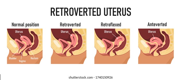
Diagram of uterus and bladder. Genital System Development - Embryology uterus - The female internal genital (reproductive) tract forming a hollow muscular walled organ, embryonically derived from the paramesonephric ducts. vagina - Part of female genitalia formed from the paramesonephric duct develops as a muscular tube between the uterus and vestibule. Prolapse of the Uterus, Bladder, Bowel, or Rectum - HERS Foundation Broad bands of uterine ligaments provide structural support to the uterus and pelvis. The uterine ligaments may weaken, stretch, or they can be Prolapse can also be familial, occurring in more than one woman in the same family. The uterus sits between the bladder and the bowel, supporting them... 1,209 Bladder Uterus Stock Illustrations, Cliparts and Royalty Free... Fistula between bladder and uterus or and rectum and uterus caused by prolonged labour in a pregnant woman. Beautiful woman doctor with pointer and presentation board with diagram of human anatomy space flat illustration. Positions of uterus stock vector. Illustration of anteverted - 35980715 Diagram for variants of uterine position. Normal uterus rests on the superior surface of the empty bladder. Normal Uterus Positions: Anteflexed and Anteverted.
Uterus - Wikipedia The uterus (from Latin "uterus", plural uteri) or womb (/wuːm/) is a major female hormone-responsive secondary sex organ of the reproductive system in humans and most other mammals. Uterus - human anatomy organs | UTERUS DIAGRAM The walls of the uterus provide a lining that is suitable for implantation. Prenatal development follows a course of approximately nine months which provides Positioned near the lower division of the pelvic cavity, the uterus is in close proximity to the bladder, and the rectum sandwiched in between the two. Histology slides and handmade diagrams histological diagram of uterus in menstrual phase. histological diagram of transverse section of urin... gall bladder histology slides. Duodenum histology slide. Histological Diagram of Uterus | Histology | UHS - YouTube Lecture On Microscopic Structure Of Uterus. Histology of uterus. Medicos World.
Bladder | Radiology Key 2.1 Normal Bladder Anatomy. The bladder consists of two main parts: (1) the body where urine is collected, and (2) the bladder neck, which is a 2-3 cm long, funnel-shaped extension of the body that traverses the urogenital diaphragm and connects with the urethra. Pregnancy & Bladder Control: Causes, Diagnosis & Treatment Bladder control problems can happen both during pregnancy and after childbirth. Causes of bladder control issues can include pelvic organ prolapse, weakened pelvic floor muscles and damaged pelvic nerves. Diagram of uterus and bladder | Quizlet Start studying uterus and bladder. Learn vocabulary, terms and more with flashcards, games and other study tools. Only RUB 220.84/month. uterus and bladder. STUDY. Male & Female Dog Reproductive Systems — Organs and Hormones The uterus holds a pair of uterine horns that, together in unison, create the entirety of the uterus body. The uterus is then attached to the vagina The channel that serves as a basic midpoint between the uterus and the vaginal canal is the part of the female sexual reproductive system called the cervix.
Anterior prolapse (cystocele) - Symptoms and causes - Mayo Clinic
bladder uterus diagram | Answers from Doctors | HealthTap "is eating ice bad for bladder or uterus? no issues at all just asking beacuse i eat alot of ice." Answered by Dr. Alok Patel: Not usually... Unrelated: A retroverted uterus would actually be pointing away from your bladder, towards your back/buttocks. So it is unlikely to be the cause of your bladder ...

Basic Anatomy: The Urinary System | Embarrassing Problems
To the question of syntopic relations between the... In the sectional material with use of anatomical research methods were studied projection-syntopic relationship with vaginal fornix of vesico-uterine deepening and subperitoneal structures, located between the bladder and the uterus.

Uterine Position Normal Uterus Rests On Stock Illustration ...
Female reproductive system Notes: Diagrams & Illustrations | Osmosis 2 OSMOSIS.ORG Uterus ▪ Located posterior to bladder, anterior to rectum ▪ Fundus (top) → uterine body → uterine isthmus → cervix (neck of uterus) ▫ Cervical Chapter 8 Reproductive System Figure 8.7 Anterior view of the uterus and lateral view of the uterus in relationship to surrounding structures.
Urotrauma: Symptoms, Diagnosis & Treatment - Urology Care ...
Pathology Outlines - Anatomy & histology | Uterus Uterine cervix: lower one - third of uterus, which attaches to vaginal canal; see Histology. Fundus: domed superior portion of uterus located superior to points of fallopian tube insertion. Anterior portion can be identified by a higher peritoneal reflection, due to location of the bladder in vivo ( J Clin...

Rectocele picture | Pelvic organ prolapse, Uterine prolapse ...
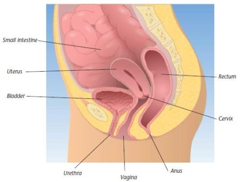
Female reproductive organs: Anatomy and functions | Kenhub Female anatomy diagram: Uterus and ovaries. The uterus is divided into three parts: Body (corpus) - the main part of the uterus, connected to the uterine As it reflects from the uterus to the rectum and urinary bladder, two folds are formed: the rectouterine pouch (of Douglas) and the vesicouterine...
Types Pelvic Organ Prolapse Cystocele Uterine Stock Vector ...
26 Diagram Of Uterus And Bladder - Wiring Database 2020 Uterus Ovaries And Bladder Posterior Carry All Pouch. The upper and side surfaces of the bladder are covered by peritoneum also called serosa. Diagram of uterus and bladder. Uterine prolapse can occur in women of any age. The uterus and the vagina. Outer surfaces of the bladder.

Anatomical structure of the female pelvis and the ...
Uterus Diagram High Res Illustrations - Getty Images Select from premium Uterus Diagram images of the highest quality. diagram of woman's pelvis and curled up foetus. - uterus diagram stock illustrations. female silhouette with simplified kidneys, bladder, ovaries and uterus. - uterus diagram stock illustrations.
Pelvic Organ Prolapse - Idaho Falls Care and Treatment
Uterus | Radiology Reference Article | Radiopaedia.org The uterus is an extraperitoneal hollow, thick-walled, muscular organ of the female reproductive tract that lies in the lesser pelvis. The uterus is divisible into two portions: body and cervix. About midway between the apex and base is a slight constriction known as the isthmus.
UTERUS
Diagram Of Uterus And Bladder - Wiring Diagram Source Bladder diagram uterus image via humanbodyanatomyco. The uterus is supported by several ligaments including the round ligament attaches to the uterus near the junction of the uterine tube and runs through the inguinal canal to the labia majora the ligament of the ovary attaches to the uterus...
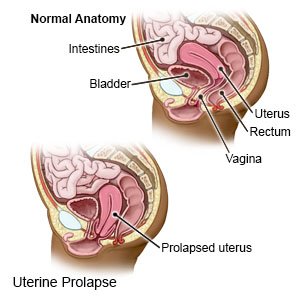
Uterine Prolapse - What You Need to Know
Uterus , Parts, position, supports, arterial supply and lymphatic... Uterus anatomy - parts, position, ligaments, arterial spply and lymphatic drainage. simple easy notes on uterus for quick revision before exams. Angle of anteflexion: The body of uterus is slightly bent forwards on the cervix and the angle between the long axis of body of uterus and the long axis of...
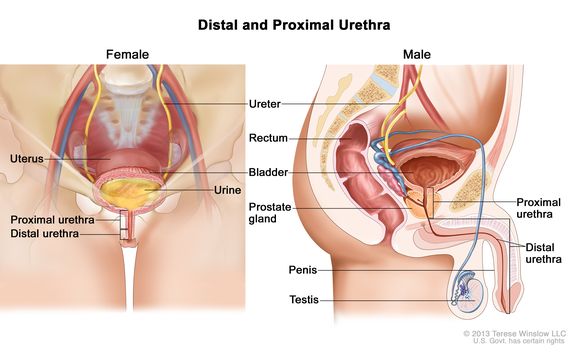
Definition of urethra - NCI Dictionary of Cancer Terms ...
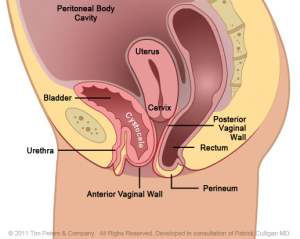
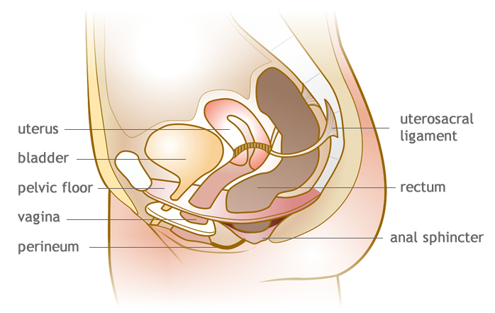
The Anatomy of Pelvic Support - Atlas of Vaginal Reconstructive... Diagram of the pelvic support, depicting the bladder, the obturator muscle and foramen, and the arcus tendineus where the levators are inserting. The sacrouterine ligaments provide the most important support to the uterus and the vaginal cuff (see Fig. 1.3). The ligaments anchor the cervix to the...
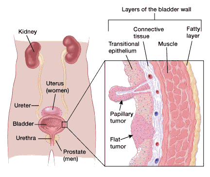
What Is Bladder Cancer?
Ultrasound Scanning Planes - Sonographic Findings 16 Reverberations: Longitudinal scan of the uterus and bladder (B) with. 35 Diagram of the arterial vessels arising from the aorta and the tributaries of the vena cava. These vessels can be distinguished sonographically and can provide useful landmarks for intra-abdominal scanning.

Drawing of female pelvis (midsagittal view) shows the anatomy ...
Intraoperative view of uterus, bladder, excised VUF tract, and... ... Vesicouterine fistula (VUF) is a rare abnormal communication between the bladder and the uterus, accounting for approximately 1%-4% of the genitourinary fistulas. [1] It was first described in 1957 by Youssef, who defined the classic triad of presentation symptoms: vaginal urine leakage, amenorrhea...
UTERUS
The Uterus - Structure - Location - Vasculature - TeachMeAnatomy Cervix - lower part of uterus linking it with the vagina. This part is structurally and functionally different to the rest of the uterus. See here for more Anteflexed: Flexed, towards the anterior surface of the body. Thus, the uterus normally lies immediately posterosuperior to the bladder, and anterior to the...
An illustrated cross-sectional view of the uterus and bladder ...
Urinary Bladder - Earth's Lab Urinary Bladder is one of the organs of the human body located in the anterior part of the pelvis, which is also a muscular Urinary bladder is tetrahedral in shape when empty and oval in shape when distended. Capacity. In the female, it's split from the cervix of uterus and by the vesicouterine pouch.
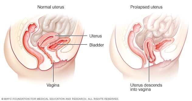
Uterine prolapse - Symptoms and causes - Mayo Clinic

Uterus | Radiology Reference Article | Radiopaedia.org
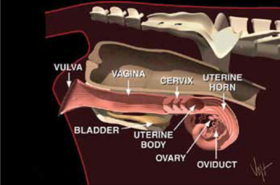
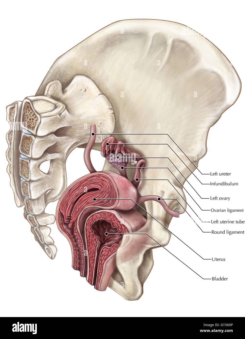
Reproductive Anatomy and Physiology of Cattle

1,972 Bladder Uterus Stock Photos and Images - 123RF

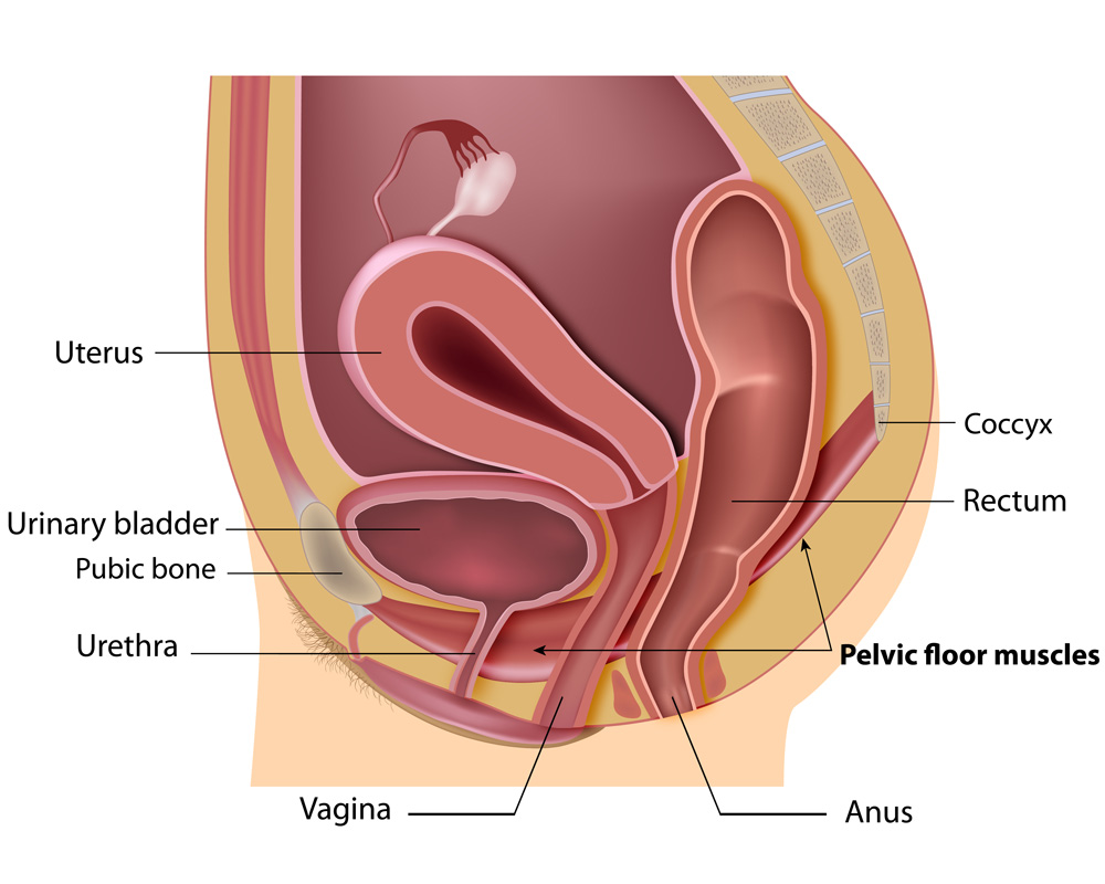
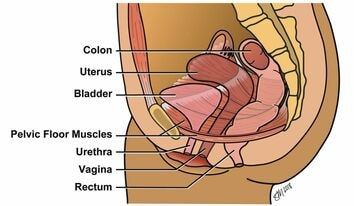
Pelvic Floor Muscles - Its Functions more | Elara Care
5: Positions of uterus: Normal position of the uterus is ...
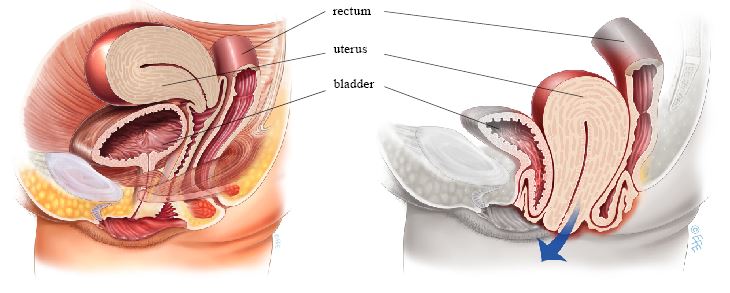
Vaginal Hysterectomy for Prolapse - Your Pelvic Floor
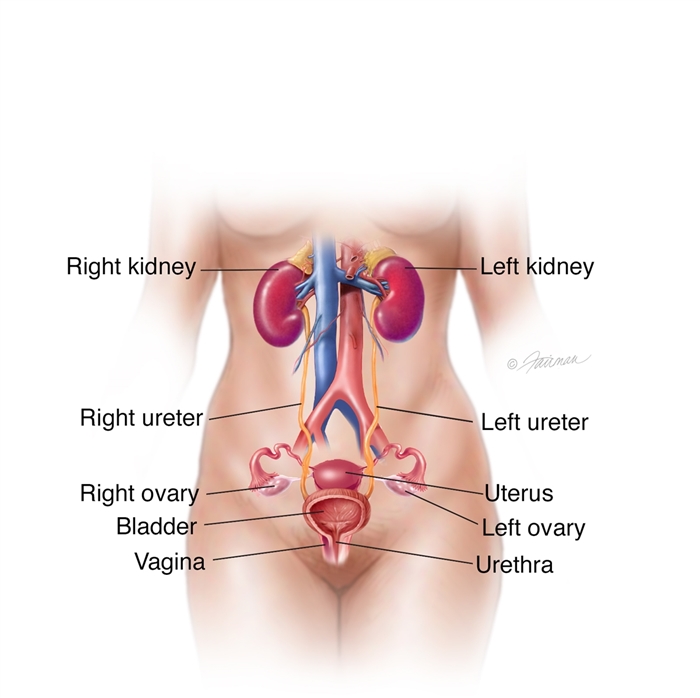
:max_bytes(150000):strip_icc()/FemaleUrinarySystemKocakayaaliGetty-8bc0982046934d24a9cfcf4f7c29ac1c.jpg)
Female Body Diagram: Parts of a Vagina, Location, Function
Cystocele Treatment - Gynecologic & Reconstructive Surgery
Female pelvic area with labels for the cervix, vagina ...

The Female Hip and Pelvis | Musculoskeletal Key
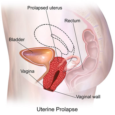
Uterine Prolapse - Physiopedia

For patients | pfm medical ag
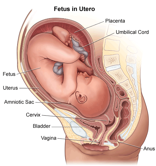
Anatomy: Fetus in Utero
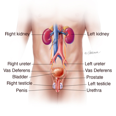
The Urinary Bladder - Structure - Function - Nerves ...
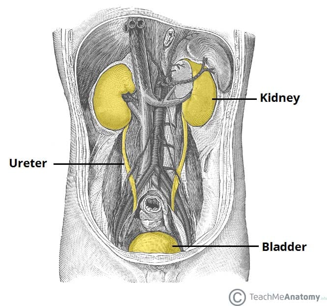
Extrinsic Obstruction of the Ureter: Symptoms, Diagnosis ...
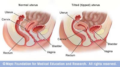
Tipped (tilted) uterus - Mayo Clinic
Pelvic Anatomy
Urogynecology: About Pelvic Organ Prolapse :: Minnesota ...

1,559 Uterus Diagram Stock Photos, Pictures & Royalty-Free ...

Urinary bladder - Wikipedia
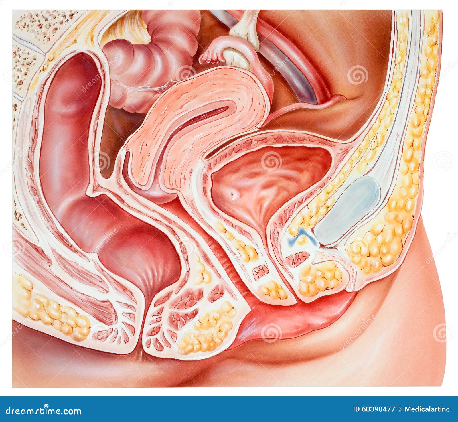
Pelvis - Female, Anatomy stock image. Image of dorsal - 60390477

Female pelvic floor 1: anatomy and pathophysiology | Nursing ...

Doctors Found A Glass Tumbler Was Cause Of A Woman's UTI Symptoms
Pelvic Organ Prolapse

Prolapse
0 Response to "41 diagram of uterus and bladder"
Post a Comment