40 diagram of eye muscles
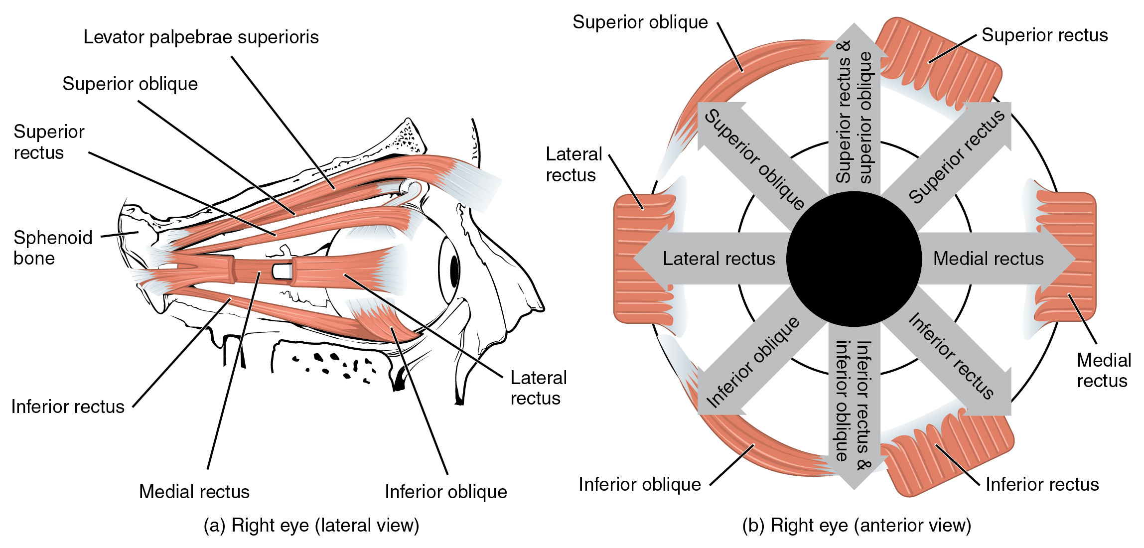
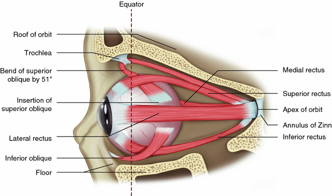
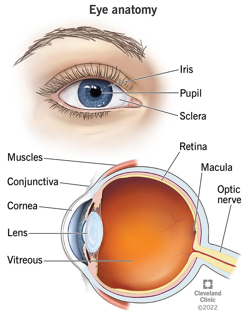
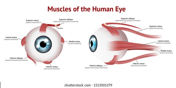
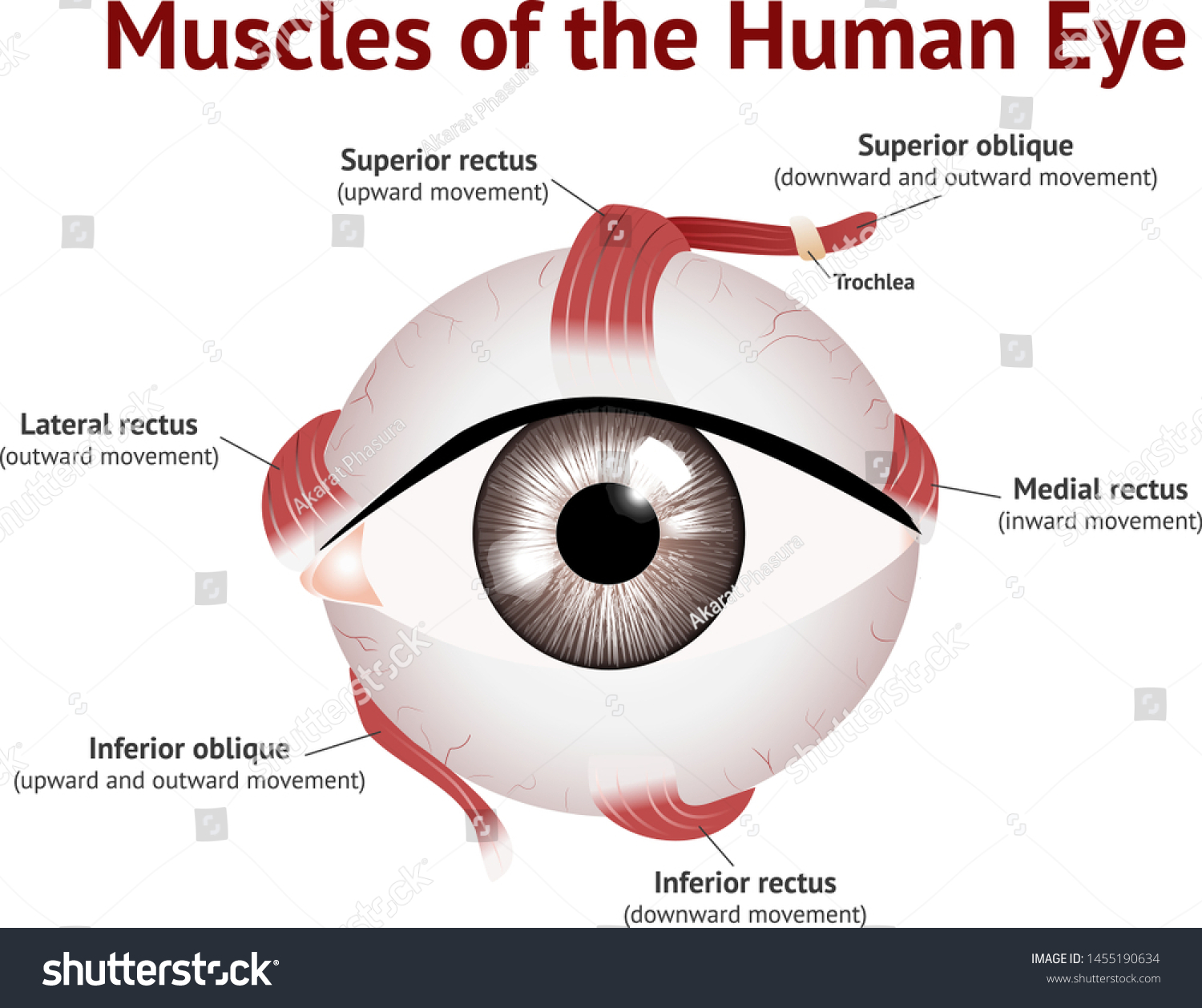
diagram of eye muscles - Graph Diagram This human anatomy diagram with labels depicts and explains the details and or parts of the Diagram Of Eye Muscles. Human anatomy diagrams and charts show internal organs, body systems, cells, conditions, sickness and symptoms information and/or tips to ensure one lives in good health. Chapter 4: Eye movements | Extraocular muscle function All eye muscles have a resting muscle tone that is designed to stabilize eye position. During movements, certain muscles increase their activity Sweating is depressed in the face on the side of the denervation, the upper eyelid becomes slightly ptotic and the lower lid is slightly elevated due to...
Sclera Anatomy, Function & Diagram | Body Maps 19.1.2018 · The sclera is the part of the eye commonly known as the “white.” It forms the supporting wall of the eyeball, and is continuous with the clear cornea. The sclera is covered by the conjunctiva ...

Diagram of eye muscles
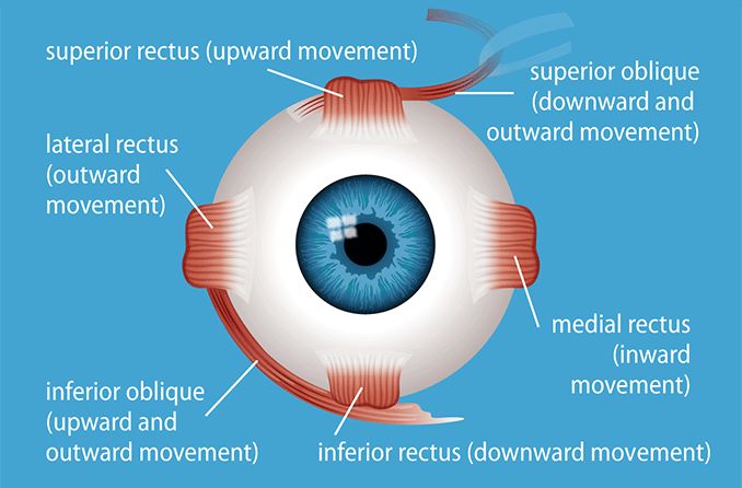
Diagram of eye muscles diagram | Quizlet Start studying eye muscles diagram. Learn vocabulary, terms and more with flashcards, games and other study tools. superior oblique. - depresses eye and turns it laterally - IV (trochlear). inferior rectus muscle. Eye anatomy: Muscles, arteries, nerves and lacrimal gland There are two groups of eye muscles: ... Six extraocular muscles move the eye: superior rectus, inferior rectus, medial rectus, lateral rectus, superior oblique ... Vision and Eye Diagram: How We See "The eye is a container," says Richard Rosen, M.D., a vitreoretinal surgeon at the New York Eye and Ear Infirmary of Mount Sinai, in New York City. Small elastic muscles, known as ciliary muscles, which are attached to the lens, help it change its shape in order to focus at various distances.
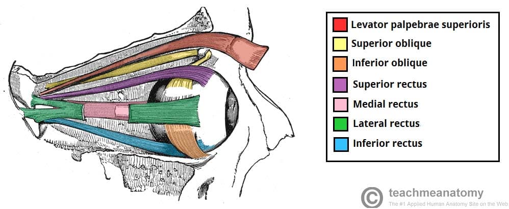
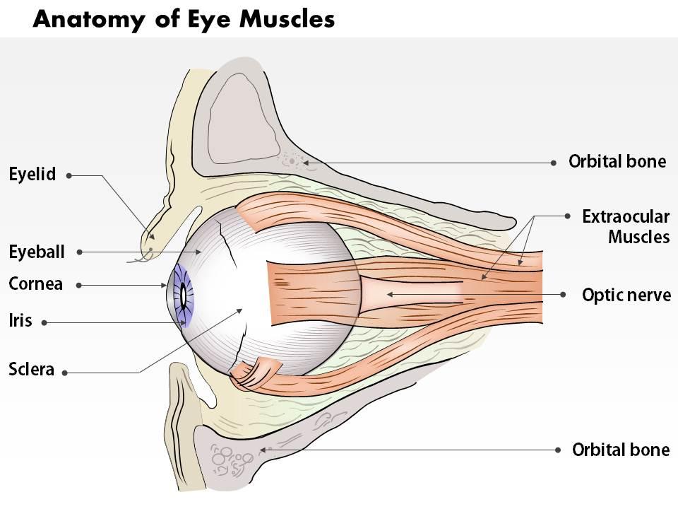
Diagram of eye muscles. The Human Eye (Eyeball) Diagram, Parts and Pictures ... The six extrocular muscles move the eyelids and eyeballs. Levator palpebrae superioris – lifts the upper eyelid Superior oblique – moves eyeball to the outer side (abduct) and downwards (depresses), medial rotation Inferior oblique – moves eyeball to the outer side (abduct) and upwards (elevates), lateral rotation Human eye - Wikipedia Schematic diagram of the human eye. It shows a horizontal section through the right eye. The movements of the eye are controlled by six muscles attached to each eye, and allow the eye to elevate, depress, converge, diverge and roll. Muscles of the Eye Six skeletal muscles surround the eye and control the many diverse movements of the eyes. These muscles, although small and not particularly strong, are exceptionally fast and precise. They allow the eye to perform many complex tasks, including tracking moving objects, scanning for objects, and... Eye muscle exercise - Physiopedia Original Editor - Aarti Sareen Top Contributors - Khloud Shreif , Aarti Sareen , Kim Jackson and Lucinda hampton. The eye divided into three layers; the outermost layer is a fibrous layer and it is consists of the cornea that is transparent located at the center of the eye...
The Human Eye - Diagram, Parts, Working, Function and Work ... Six muscles are in the eye. They are responsible for controlling the movement of the eye. The most common kinds of muscles that are in the eye are the lateral rectus, medial rectus, inferior oblique, or superior rectus. (Image will be Uploaded soon) Parts of the Human Eye. Pupil: The pupil is a small opening in the iris. Extraocular muscles: Anatomy and movements | Kenhub Extraocular muscles are also referred to as the extrinsic (arising externally) or muscles of the orbit. There are 6 of these extraocular muscles that control eye movement (cows only have 4 of these), and one muscle that controls eyelid elevation. The position of the eye at the time of muscle contraction is... Eyes | ORBICULARIS OCULI MUSCLE The eyelid muscles also control the blink reflexes. Blinking provides moisture to the eyes and the cornea by using tears (produced in the tear glands) and oily substances (secreted Hence, there is a cross-over correlation from the brain to the organ (see GNM diagram showing the motor homunculus). Vision and the eye's anatomy | HealthEngine Blog Extraocular muscles (muscles outside the eye) allow the eye to move within its orbit. Six of these eyeball muscles attach to each eye. The following diagram illustrates the events that occur in a photoreceptor in response to light, initiating an action potential in the visual pathway.
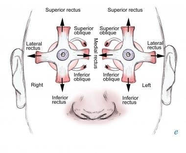
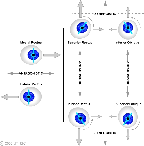
PDF FIGURE 2-12. Diagram of the superior oblique (SO) muscle and... FIGURE 2-9. Diagram of the eye and orbit from a top view looking down on the superior rectus (SR) muscle. FIGURE 2-14. Diagram of posterior anatomy of the eye and muscles. Note the proximity of the inferior oblique to the macula and vortex veins (vv). Eye Diseases: Symptoms & Causes of 19 Common Eye Problems Lazy Eye. Lazy eye, or amblyopia, happens when one eye doesn’t develop properly.Vision is weaker in that eye, and it tends to move “lazily” around while the other eye stays put. It’s found ... The Anatomy of the Human Eye - with diagram of the eye. This simple introduction the subjects of 'the eye' and 'visual optics' includes a simple diagram of the eye together with definitions of the parts of the The ciliary muscle is a ring-shaped muscle attached to the iris. It is important because contraction and relaxation of the ciliary muscle controls the shape... Extraocular Muscles | Eye Movement | Eye Muscles | Geeky Medics The extraocular muscles (EOM) are responsible for controlling the movements of the eyeball and upper eyelid. AO = All other extraocular muscles: innervated by the third nerve. Figure-2. Schematic diagram of the actions of the extraocular muscles and their innervation.
The Anatomy of Human Eye with Diagram | EdrawMax Online The human eye diagram is a visual depiction of the human eye. The following aspects are essential when constructing a human eye diagram. One muscle inside the iris constricts the Pupil in bright light, for example, full sunlight, and another iris muscle dilates the Pupil in dim lighting and darkness.
Anatomy of the eye | Eye Structure Diagram | Patient The movement of each eye is controlled by six muscles that pull the globe of the eye in various directions. They work together in a synchronised way. Eyelids help to spread the tear film across the eye by blinking. They also produce a special oil which slows down the evaporation of the tear film.
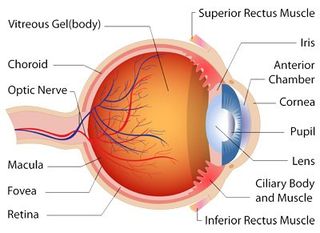
Eye Anatomy Handout - National Eye Institute Eye Anatomy Handout Author: National Eye Institute , National Eye Health Education Program Subject: Diabetes and Healthy Eyes Toolkit and Website Keywords: Eye anatomy, eye diagram, cornea, iris, lens, macula, optic nerve, pupil, retina, vitrous gel, diabetic eye disease. Created Date: 6/27/2012 11:57:40 AM
Schematic Diagram of the Human Eye: Structure of the eye and... The eye has six muscles which control the eye movement, all providing different tension and torque. The eye works a lot like a camera, the pupil provides the f-stop, the iris the aperture stop, the cornea resembles a lens. The way that the image is formed is much like the way a convex lens forms an image.
Parts of the Eye - National Eye Institute Eye Diagram Handout Author: National Eye Health Education Program of the National Eye Institute, National Institutes of Health Subject: Handout illustrating parts of the eye Keywords: parts of the eye, eye diagram, vitreous gel, iris, cornea, pupil, lens, optic nerve, macula, retina Created Date: 12/16/2011 12:39:09 PM
Anatomy of the Eye - American Association for Pediatric... These muscles originate in the eye socket (orbit) and work to move the eye up, down, side to side, and rotate the eye. The inferior oblique is an extraocular muscle that arises in the front of the orbit near the nose. It then travels outward and backward in the orbit before attaching to the bottom part of the...
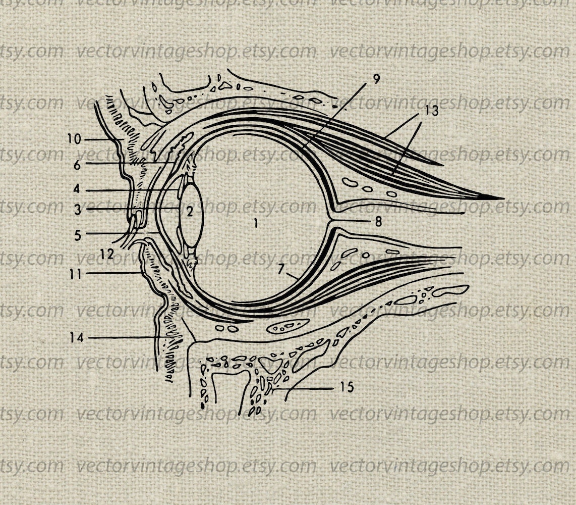
EYE ANATOMY SVG File, Eye Muscles Diagram, Medical Vector Clip Art, Science Vintage Illustration, Medical Art Commercial Use, png jpg eps
Structure and Function of the Human Eye Human eyes are "camera-type eyes," which means they work like camera lenses focusing light onto film. The cornea and lens of the eye are analogous to the Light enters the eye by passing through the transparent cornea and aqueous humor. The iris controls the size of the pupil, which is the opening...
Eye Pictures, Anatomy & Diagram | Body Maps Jan 28, 2015 · A series of muscles helps the eye move. The first set is the superior and inferior rectus muscles, which allow upward and downward motion. The medial and lateral rectus muscles allow the eye to...
Structure of Human Eye (With Diagram) | Human Body The ciliary muscles are smooth muscles and are of two types: circular and meridional. Attached to the ciliary body are the suspensory ligaments, which are in turn attached to the capsule that surrounds the lens of the eye. Related Articles: Human Eye: Structure of Human Eye (With Diagram) | Biology.
Eye Muscles | Vivid Vision The muscles of the eye are designed to stabilize and move both eyes. Special nervous centers located throughout the brain and brainstem interact with each muscle pair (right and left) to coordinate precise movements of the eyes with limited conscious input.
Extraocular Muscle Actions: Eye Movements, Rectus Muscles... Yoke muscles are the primary muscles in each eye that accomplish a given version (eg, for right gaze, the right lateral rectus and left medial rectus muscles). Each extraocular muscle has a yoke muscle in the opposite eye to accomplish versions into each gaze position.
Diagram of extraocular muscles Extraocular Muscles. Master Diagram · Eye Movements · Table of Contents · Subject Index · Table of Contents [When not using framtes]
Actions of the ocular muscles - YouTube Actions of the ocular muscles. Смотреть позже.
Eye muscles - All About Vision Eye Diagram. Extrinsic eye muscles (also called extraocular muscles) are attached to the outside of the eyeball and enable the eyes to move in all directions of sight. Additionally, a muscle called the levator palpebrae superioris (LPS) raises the upper eyelid and keeps it in position.
Human Eye Ball Anatomy & Physiology Diagram The eye is cushioned within the orbit by pads of fat. In addition to the eyeball itself, the orbit contains the muscles that move the eye, blood vessels, and The eyelids serve to protect the eye from foreign matter, such as dust, dirt, and other debris, as well as bright light that might damage the eye.
How Your Eyes Work - YouTube Your eyes see, but how does vision happen? Find out how the eyes and brain work together in this eye video.
Eye muscles - Owlcation Muscles of the eye are very strong and efficient, they work together to move the eyeball in many different directions. The main muscles of the eye In the diagram above - anatomy of the eye, the artery is shown in red while the vein is shown in blue. Tear Duct. This is a small tube that runs from...
Eye Anatomy: Parts of the Eye and How We See - American Academy... The eye has many parts, including the cornea, pupil, lens, sclera, conjunctiva and more. They all work together to help us see clearly. The surface of the eye and the inner surface of the eyelids are covered with a clear membrane called the conjunctiva.
The Extraocular Muscles - The Eyelid - Eye... - TeachMeAnatomy The extraocular muscles are located within the orbit, but are extrinsic and separate from the eyeball itself. They act to control the movements of the eyeball and the superior eyelid.
Human Eye: Anatomy, parts and structure - Online Biology Notes Four sets of these muscles are straight muscles; superior, inferior, medial and lateral rectal muscle and two sets are oblique muscles; superior and inferior oblique It is muscular, pigmented and opaque diaphragm which hangs in the eye ball in front of lens. It has small circular opening called pupil.
human eye | Definition, Anatomy, Diagram, Function ... human eye, in humans, specialized sense organ capable of receiving visual images, which are then carried to the brain. The eye is protected from mechanical injury by being enclosed in a socket, or orbit, which is made up of portions of several of the bones of the skull to form a four-sided pyramid, the apex of which points back into the head.
Vision and Eye Diagram: How We See "The eye is a container," says Richard Rosen, M.D., a vitreoretinal surgeon at the New York Eye and Ear Infirmary of Mount Sinai, in New York City. Small elastic muscles, known as ciliary muscles, which are attached to the lens, help it change its shape in order to focus at various distances.
Eye anatomy: Muscles, arteries, nerves and lacrimal gland There are two groups of eye muscles: ... Six extraocular muscles move the eye: superior rectus, inferior rectus, medial rectus, lateral rectus, superior oblique ...
Diagram of eye muscles diagram | Quizlet Start studying eye muscles diagram. Learn vocabulary, terms and more with flashcards, games and other study tools. superior oblique. - depresses eye and turns it laterally - IV (trochlear). inferior rectus muscle.













0 Response to "40 diagram of eye muscles"
Post a Comment