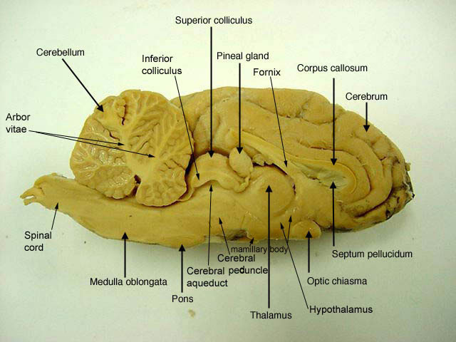
37 sheep brain diagram labeled
DISSECTION OF THE SHEEP'S BRAIN Introduction The purpose of the sheep brain dissection is to familiarize you with the three-dimensional structure of the brain and teach you one of the great methods of studying the brain: looking at its structure. One of the great truths of studying biology is the saying that "anatomy precedes physiology". neuroanatomy by examining the brain of the sheep. We will be looking as several structures within the central nervous system (CNS), which is composed of the brain and spinal cord. You will learn individual structures now; later you will be looking at brain systems to see how different parts of the brain form functional units.
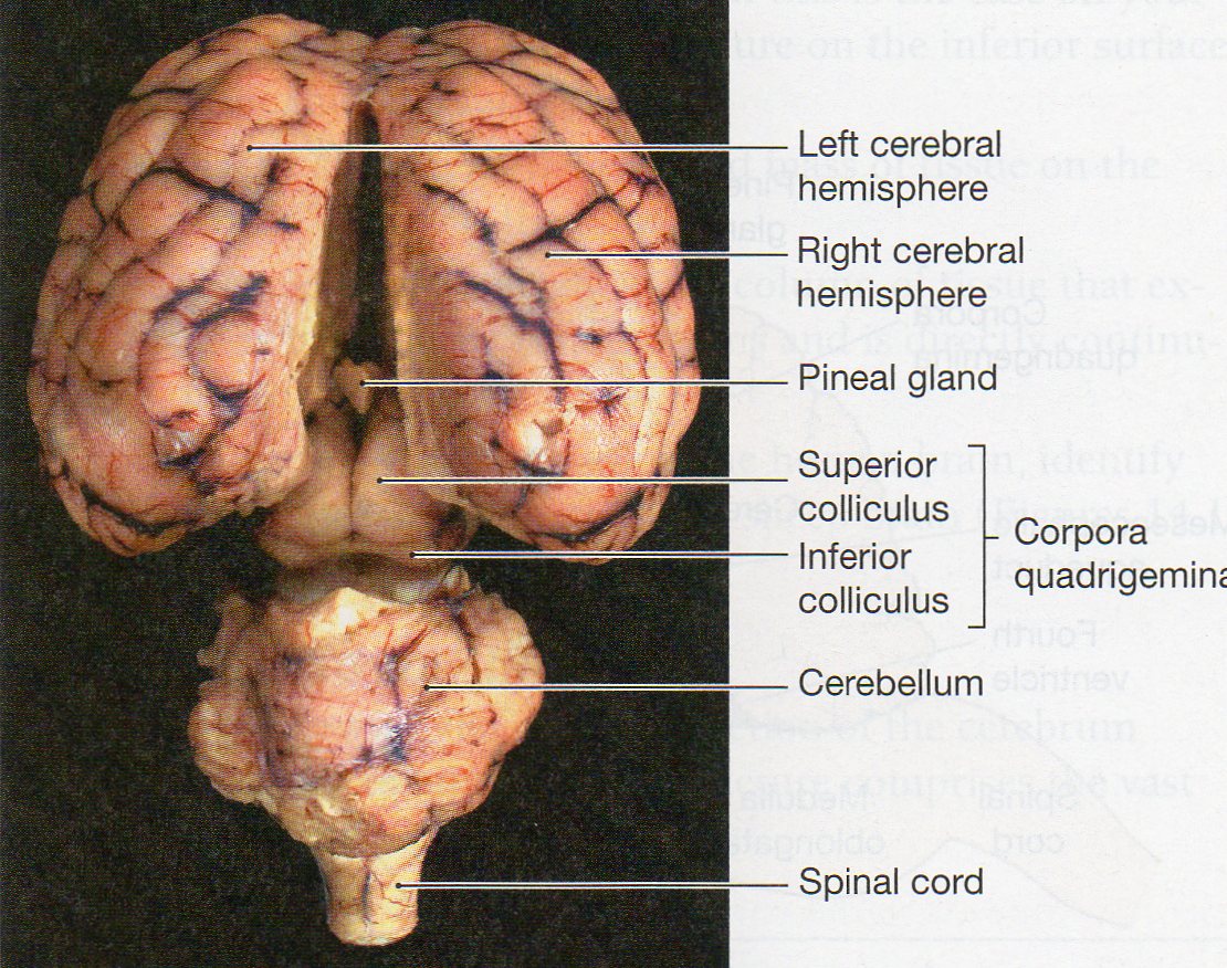
The sheep brain is quite similar to the human brain except for proportion. The sheep has a smaller cerebrum. Also, the sheep brain is oriented anterior to ...

Sheep brain diagram labeled
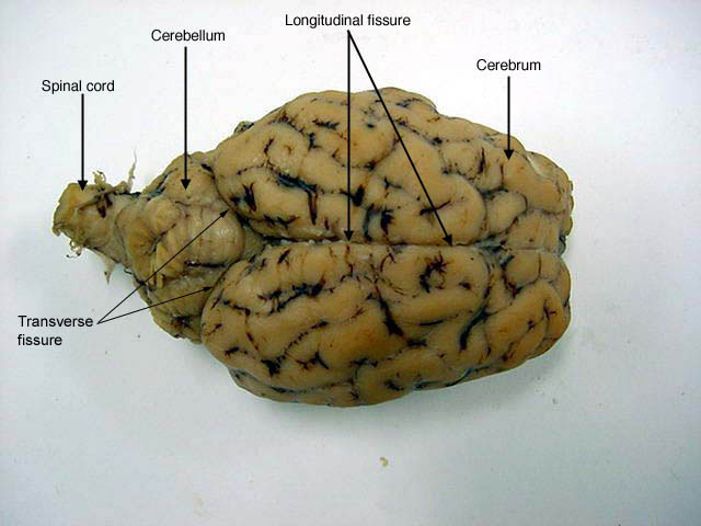
External and Midline Anatomy. Examine the sheep brain with the membranes intact. You should be able to identify and use the following directional terms:.14 pages The sheep brain is exposed and each of the structures are labeled and described in a sequential manner, in the same way that a real dissection would occur. Sheep Brain Dissection with Labeled Images Sheep Brain Dissection 1. The sheep brain is enclosed in a tough outer covering called the dura mater. 5 3 11 6 22 16 18 1. Gray Matter 2. White Matter 3. Corpus Callosum 4. Lateral Ventricle 5. Caudate Nucleus 6. Septum Pellucidum 7. Fornix 8.
Sheep brain diagram labeled. Label theBrain of the Sheep. Publisher: Biologycorner.com; follow on Google+ This work is licensed under a Creative Commons Attribution-NonCommercial 3.0 Unported License. Brain Label Answer Key. Image adapted from a photograph of the sheep brain. ... Jun 6, 2018 - A virtual sheep brain dissection guides anatomy studies with photos & blank diagrams. ... Pretty good picture of the sheep brain labeled. Sheep Brain Dissection Lab Companion In 2021 Brain Anatomy Anatomy And Physiology Brain. Sheep Brain External View Labeled Anatomia Veterinaria Anatomia Veterinaria. Diagram Of The Brain Pictagram Brain Diagram Dissection Brain Anatomy. Biology 02 Steiner Flashcards Sheep Brain Studyblue. Sheep Brain Dissection Project Guide Hst Learning Center ... Sheep Brain Anatomy Lab Manual. Based on original material by R. N. Leaton, Dartmouth College. Contributors to this version: Al Sorenson, Lisa Raskin, Sarah Turgeon, Steve George, and JP Baird. I. Introduction. The brain of the sheep is useful for study because its anatomy is similar to human brain anatomy. Although exact proportions (and names ...
8 Jul 2021 — Anatomy & Physiology one organ at a time...Sheep Brain Dissection may be downloaded from https://www.biologycorner.com/anatomy/chap9.html. Sheep Brain Dissection: Internal Anatomy 1. Identify the following structures on the brain and quiz each other on their functions. 2. Use the diagram on the next page to help identifying the structures. 1. Frontal Lobe 2. Temporal Lobe 3. Occipital Lobe 4. Brainstem 5. Midbrain 6. Pons 7. Medulla 8. Cerebellum 9. Cerebrum 10.Corpus callosum 11 ... Start studying Sheep Brain Dissection labeled. Learn vocabulary, terms, and more with flashcards, games, and other study tools. Sheep Brain Labeled Diagram. angelo. August 10, 2021. Labeled Brain Brain Anatomy Anatomy And Physiology Anatomy. Image Result For Sheep Brain Labeled Nursing Study Guide Anatomy And Physiology Nursing Study In 2021 Nursing Study Guide Nursing Study Human Brain Anatomy.
Sheep Neuroanatomy Lab- Labeling Worksheet. Psychology 2315- Brain and Behaviour. Kwantlen Polytechnic University. Figure 1: Dorsal view.4 pages NERVOUS SYSTEM - SHEEP BRAIN IMAGES. Sheep Brain Unlabeled. Sheep Brain in Dura Mater · Dorsal Sheep Brain · Dorsal-Colliculi ... Sheep Brain Labeled. 5 3 11 6 22 16 18 1. Gray Matter 2. White Matter 3. Corpus Callosum 4. Lateral Ventricle 5. Caudate Nucleus 6. Septum Pellucidum 7. Fornix 8. The sheep brain is exposed and each of the structures are labeled and described in a sequential manner, in the same way that a real dissection would occur. Sheep Brain Dissection with Labeled Images Sheep Brain Dissection 1. The sheep brain is enclosed in a tough outer covering called the dura mater.
transverse fissure | Brain anatomy, Cerebral cortex ...
External and Midline Anatomy. Examine the sheep brain with the membranes intact. You should be able to identify and use the following directional terms:.14 pages

sheep brain labeled 5041004 orig - Made By Creative Label
WithLuke.com / Instagram: @WithLuke / Email for business enquiries: info@withluke.com

unlabeled brain | Dissection, Nursing study guide, Brain ...

Sheep Brain Dissection Lab Companion

Labeled sheep brain

Virtual Brain Dissection | Brain anatomy, Anatomy, Human ...

Sheep Brain
Sheep head

Sheep brain dissection | Human Anatomy and Physiology Lab ...
20 New Lateral View Of Sheep Brain Labeled

Sheep Brain Diagram Lobes - Diagram Media

Sheep Brain | Brain anatomy, Nervous system anatomy ...

Black Rabbit, Byker Farm, Ouseburn Valley, Newcastle Upon Tyne, Tyne & Wear, England.
Sheep Brain

Sheep Brain

25. Midsagittal Sheep Brain (labeled) | anatomyphyslab261 ...

anatomical brain stem labeled lab model - Google Search ...

Sagittal Section -lateral ventricle (sheep's brain) http ...

34 Label The Sheep Brain - Labels Design Ideas 2020

12 best Cerebellum images on Pinterest | Cranial nerves ...

Sheep Brain
Sheep in field

Diagram of Sheep Brain - Inferior view
Young Sheep

sheep brain labeled 5041004 orig - Made By Creative Label
Sheep Brain #2

Physiological Psychology

Sheep Brain Dissection Guide with Pictures | Secondary ...

Midsagittal Section Of The Sheep Brain Labeled - img-owls
Shepherding Livestock

Sheep Brain Dissection Guide | Medicine studies, Medical ...

On the Cutting Edge: Exploring Sheep Organs | Carolina.com

Pretty good picture of the sheep brain labeled. | Brain ...

10 Best images about Biology 100: Anatomy & Phys. on ...
Who’s following who?

30 Label The Sheep Brain - Labels Database 2020
0 Response to "37 sheep brain diagram labeled"
Post a Comment