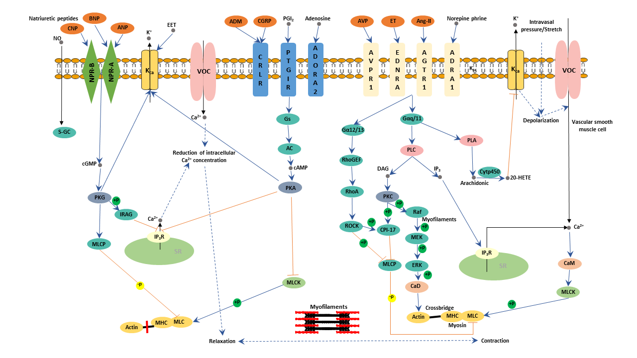
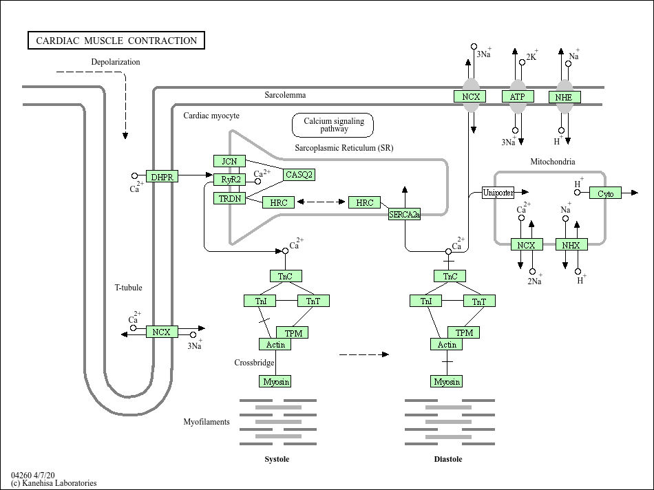
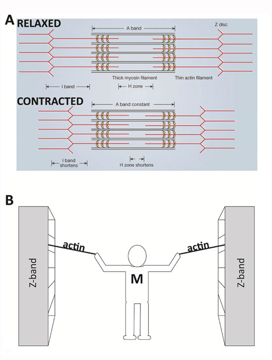
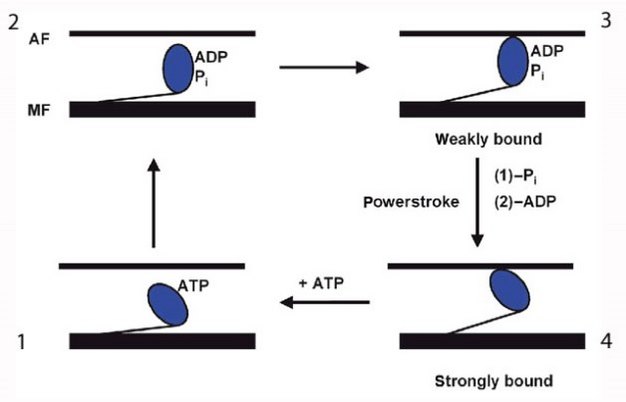
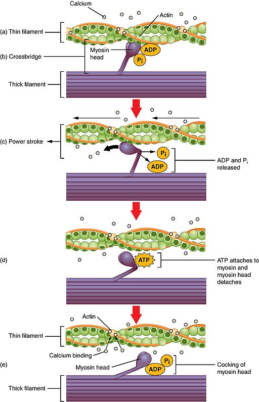
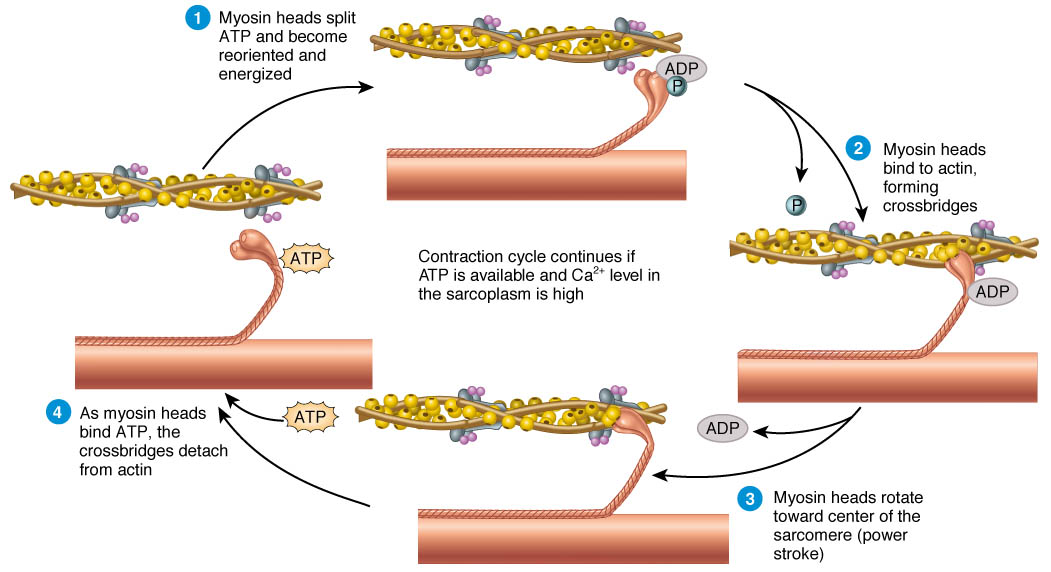
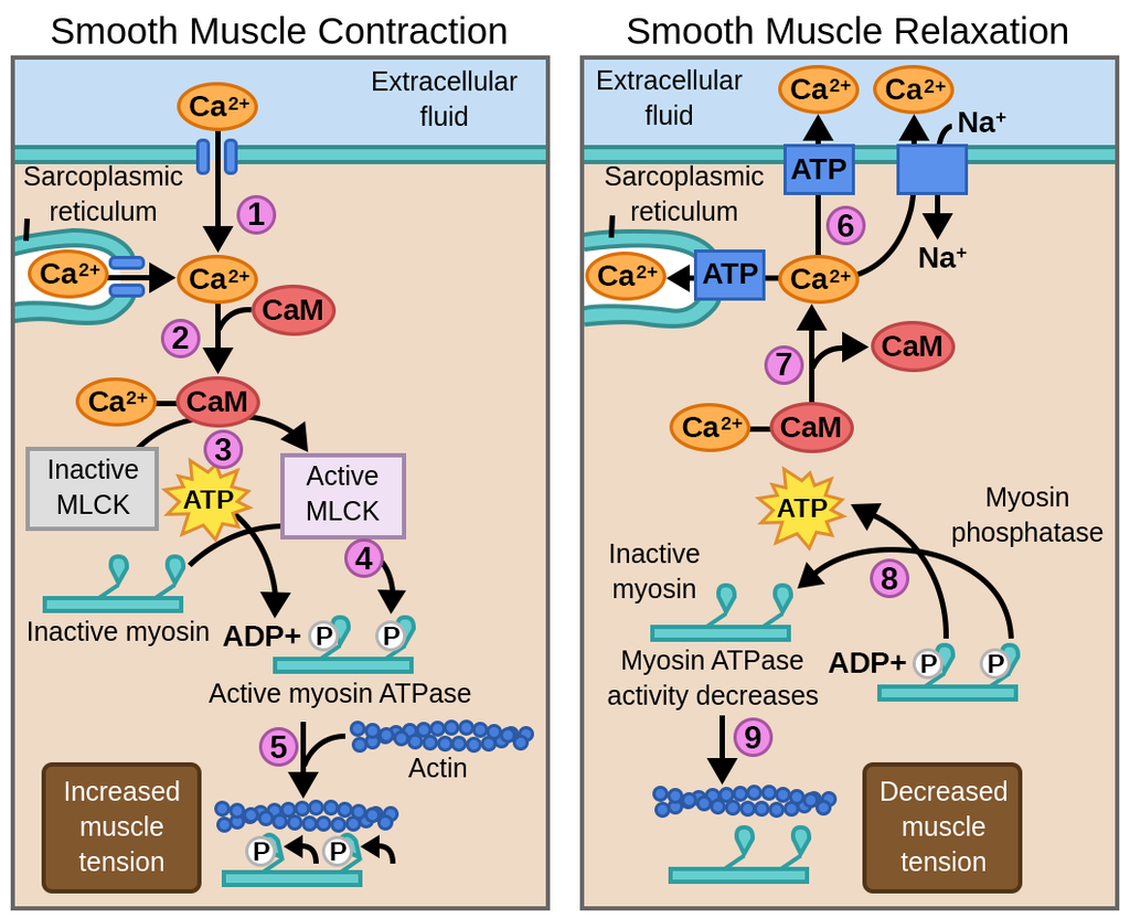
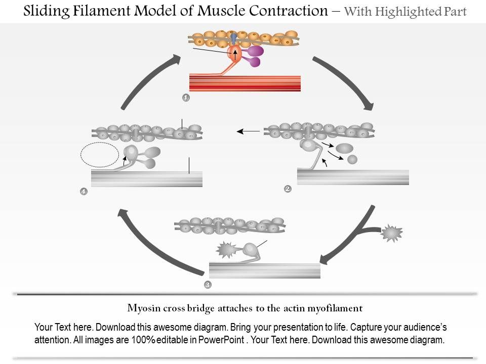
41 diagram of muscle contraction
Sliding filament model of muscle contraction. A myofibril (also known as a muscle fibril or sarcostyle) is a basic rod-like organelle of a muscle cell. Muscles are composed of tubular cells called myocytes, known as muscle fibres in striated muscle, and these cells in turn contain many chains of myofibrils. The examination was conducted by means of the isometric muscle force measurement and analysis program, 2001 Metitur Ltd. The value of maximum muscle force (Fmax) was measured and on its basis the values of maximum muscle torque (τmax) and relative muscle torque (τr) for a selected group of muscles were calculated according to:
Motor neuron and muscle contraction motor neuron the definitive guide kondo anatomy chapter 9 skeletal motor neurons in muscle contraction. Muscle Stimulation By Motor Neuron And Contraction You ... Coordination Of Independent Muscle Effectors A Conjoint Contractions Scientific Diagram

Diagram of muscle contraction
Lung adenocarcinoma (LUAD) is the most common subtype of nonsmall-cell lung cancer (NSCLC) and has a high incidence rate and mortality. The survival of LUAD patients has increased with the development of targeted therapeutics, but the prognosis of these patients is still poor. Long noncoding RNAs (lncRNAs) play an important role in the occurrence and development of LUAD. Botulinum toxin (BoNT) is a neurotoxic protein produced by the bacterium Clostridium botulinum and related species. It prevents the release of the neurotransmitter acetylcholine from axon endings at the neuromuscular junction, thus causing flaccid paralysis. The toxin causes the disease botulism.The toxin is also used commercially for medical and cosmetic purposes. The sarcomeres will stretch more if there is more blood in the chamber just before contraction. When sarcomeres are stretched further, the contraction will be equally strong. This will increase the stroke volume. If you ask the relationship between the strength of contraction and end-diastolic sarcomere length, there are two basic explanations.
Diagram of muscle contraction. Figure 1 shows the cardiac impulse conduction system, general flow of cardiac activity current, and the pattern diagram of a typical electrocardiogram waveform. P-wave and QRS-wave are waveforms which accompany the contraction of the atrium and ventricles respectively, while T-wave and U-wave are waveforms of the processes by which the ... Stimulating involuntary contractions in the digestive tract including the esophagus stomach and most of the intestines which allow food to move through the tract Vagus nerve diagram. GI digestive tract the passage through which our food travels is a tube within a tube The trunk of our body is the outer tube and the GI tract is the interior tube ... Arm Muscle On The Side Learn the muscles of the arm with unfastened quizzes, diagrams and worksheets. Start now! Tutorials and quizzes on muscles that act on the arm/humerus (arm muscle. There are one million workout splits to select from. sadly, maximum suffer from some big troubles on the way to prevent your effects, especially in case you ... Kumail Nanjiani's workout helped him pull off one of the greatest Marvel transformations of all time for Eternals. Try the muscle-building routine for yourself. Mid-2019, Kumail Nanjiani was filming the last episode of his hit HBO comedy, Silicon Valley. The show followed a group of nerdy app creators navigating the cutthroat world of big tech.
Exhalation is a passive process because muscle contraction does not occur. During exhalation, the diaphragm moves back up as the stretched lung bounces back to its normal position. As the lung returns to normal position, carbon dioxide, a waste product created by the body, moves out of the lungs through the mouth and nose. ... b Venn-diagram of all Mitocarta proteins that were up ... Phase-contrast movies of muscle contraction were acquired using the same microscope in Epi-mode and images were captured with an Andor Neo ... Figure S2 shows a schematic diagram of the CE (Rice et al., 2008), which takes in an adequate cellular machinery while maintaining an admirable balance between mechanistic detail and model parsimony. In Rice CE, to simulate the experimental protocols of internal sarcomere shortening, a series elastic element is considered to enable stretch in ... Bath application of synthetic RFa peptide to transgenic medusae following RFa + neuron ablation caused radial muscle contraction, confirming that the muscle was functionally intact (Figure S2D). Local infusion of RFa but not control peptide into the subumbrella caused local muscle contraction and margin infolding (Figure S2E).
The pelvic planes include the following: Pelvic inlet: The obstetrical conjugate is the distance between the sacral promontory and the inner pubic arch; it should measure 11.5 cm or more. The ... 40 drag the labels onto the diagram to identify the steps in a reaction both with and without enzymes. Written By Kathy W. Blatt. Tuesday, November 23, 2021 Add Comment Edit. Label the parts of the neuromuscular junction. Apr 30, 2014 · Printable worksheet and diagram on the major muscle groups in the human body for. Then, for each muscle. Muscle Group Diagram Worksheet Major Muscles. Groups of muscles usually contract todownload Muscles+Worksheet. the beginning letter of correct organ system on the blank. This page contains worksheets,. The endotoxin lipopolysaccharide (LPS) from Gram-negative bacteria exerts a direct and rapid effect on tissues. While most attention is given to the downstream actions of the immune system in response to LPS, this study focuses on the direct actions of LPS on skeletal muscle in Drosophila melanogaster. It was noted in earlier studies that the membrane potential rapidly hyperpolarizes in a dose ...
Diagram 2- Intrinsic conduction system of the heart For questions 11- 14 use Diagram 2 of the heart above and the key below. ... conduction system of heart Definition The group of cardiac muscle cells and cardiac fibres that conducts the electrical impulses or signals necessary for the contraction of the heart is known as the conduction system ...
When an impulse travels between this space, muscle contraction happens. There are around 100-500 trillion such connections in the human brain, between two nerves or nerves and glands. In this article we will discuss only the junction. Structure of Neuromuscular Junction. As we have read, the junction consists of a neuron and a skeletal muscle cell.
Muscles are active in blood circulation; it is the contraction of skeletal muscles that pushes deoxygenated blood through veins and back to the heart and lungs. Muscles are also good generators of ...
GSEA revealed that the set of KEGG genes mainly associated with VIP in AF was those involved in oxidative phosphorylation, cardiac muscle contraction, and neuroactive ligand-receptor interaction (Figure 5(b)); and the GO genes that were primarily associated with VIP were those involved in the regulation of oxidative stress-induced neuron ...
Muscle structure - muscle under the microscope. EXPLORE Unlike skeletal muscle tissue, A diagram is an example of an explanatory model. Like skeletal muscle tissue, it is striated For example, postural muscles of the neck, back, and leg have a higher proportion of type I fibres. Heart muscle (cardiac muscle) is the best example.
Well, the contraction of these muscle layers is responsible for the emptying of the bladder. That is why these muscles are known as detrusors. In between the smooth muscle layer, there present visible interstitial connective tissue. You will find a thicker circular smooth muscle fiber just above the junction of the urinary bladder with the urethra.
The sarcomeres will stretch more if there is more blood in the chamber just before contraction. When sarcomeres are stretched further, the contraction will be equally strong. This will increase the stroke volume. If you ask the relationship between the strength of contraction and end-diastolic sarcomere length, there are two basic explanations.
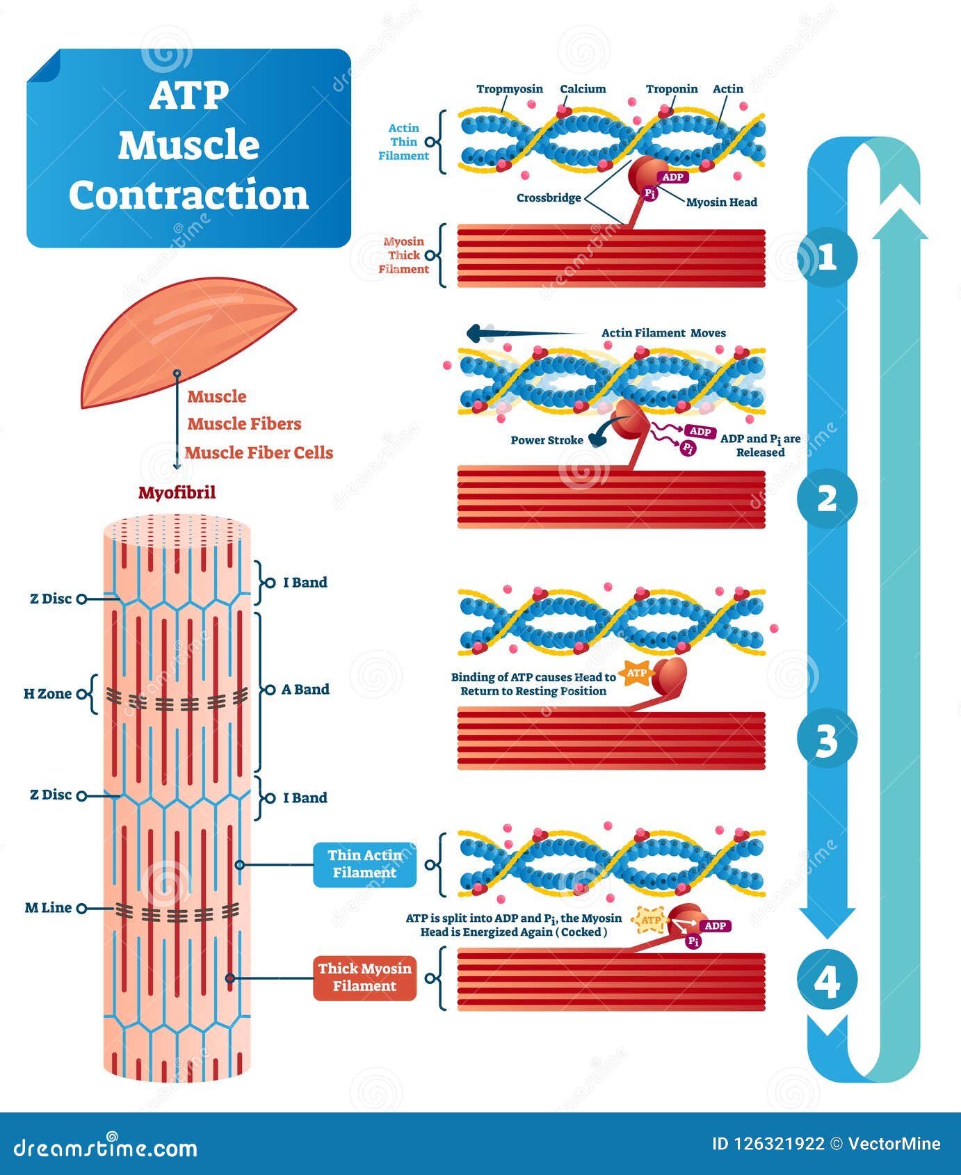
Atp Muscle Contraction Cycle Vector Illustration Labeled Educational Scheme Stock Vector Illustration Of Crossbridge Energy 126321922
Botulinum toxin (BoNT) is a neurotoxic protein produced by the bacterium Clostridium botulinum and related species. It prevents the release of the neurotransmitter acetylcholine from axon endings at the neuromuscular junction, thus causing flaccid paralysis. The toxin causes the disease botulism.The toxin is also used commercially for medical and cosmetic purposes.

Look At The Given Diagram Of Events During Muscle Contraction Named As A B C Identidy The Diagram That Shows Gradual Shortening Of Length Of Sarcomere Img Src Https D10lpgp6xz60nq Cloudfront Net Physics Images Aln Bio C15 E39 001 Q01 Png
Lung adenocarcinoma (LUAD) is the most common subtype of nonsmall-cell lung cancer (NSCLC) and has a high incidence rate and mortality. The survival of LUAD patients has increased with the development of targeted therapeutics, but the prognosis of these patients is still poor. Long noncoding RNAs (lncRNAs) play an important role in the occurrence and development of LUAD.

Skeletal Muscle A Review Of Molecular Structure And Function In Health And Disease Mukund 2020 Wires Systems Biology And Medicine Wiley Online Library

Muscle Contraction Adenosine Triphosphate Rigor Mortis Atp Hydrolysis Uterine Contraction Others Angle Text Cell Png Pngwing
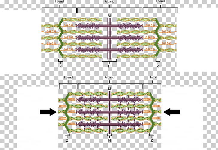
Diagram Sarcomere Myofilament Skeletal Muscle Muscle Contraction Png Clipart Actin Anatomy Angle Area Diagram Free Png













0 Response to "41 diagram of muscle contraction"
Post a Comment