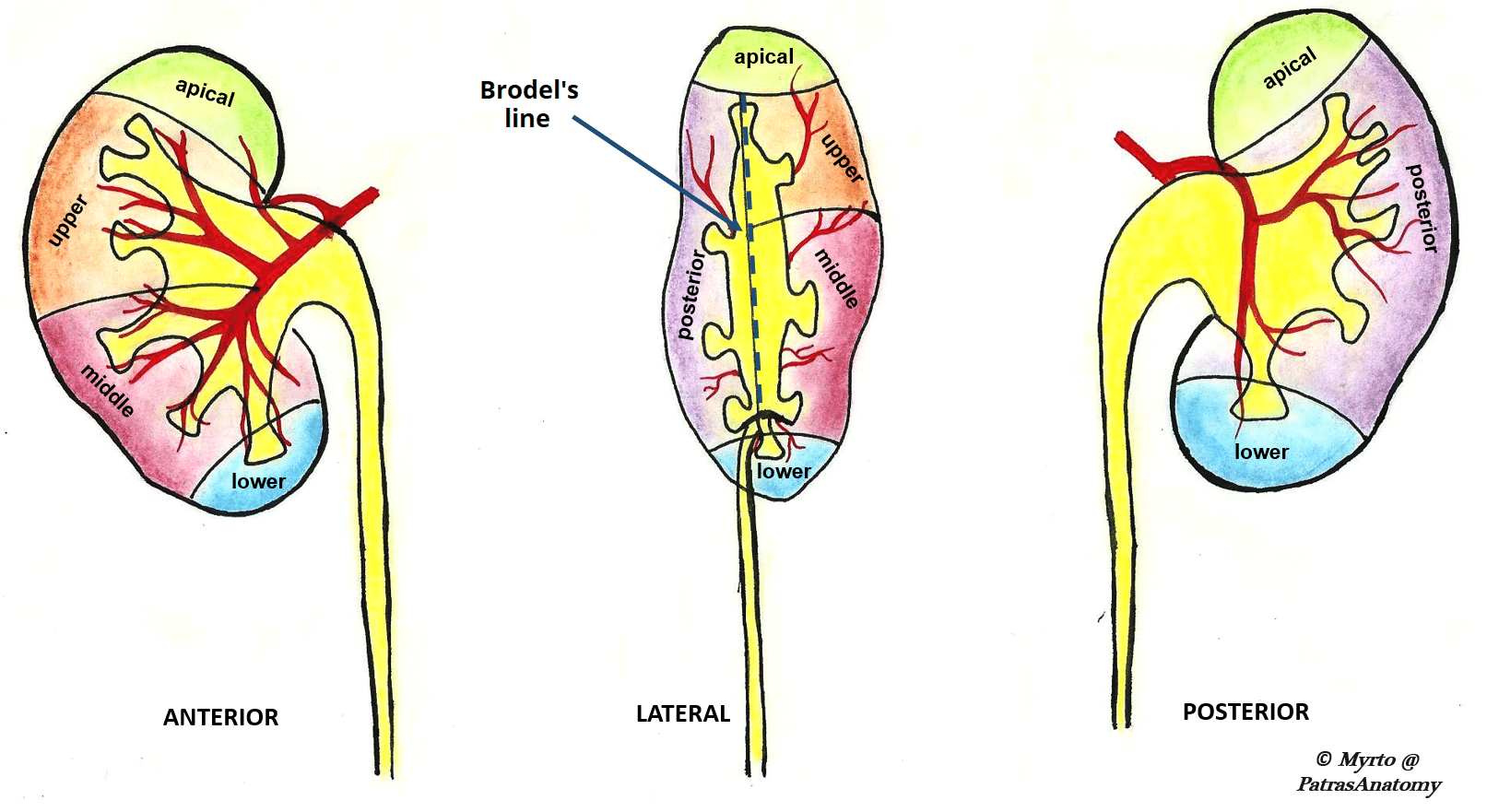
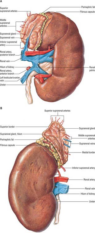
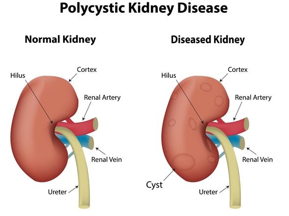
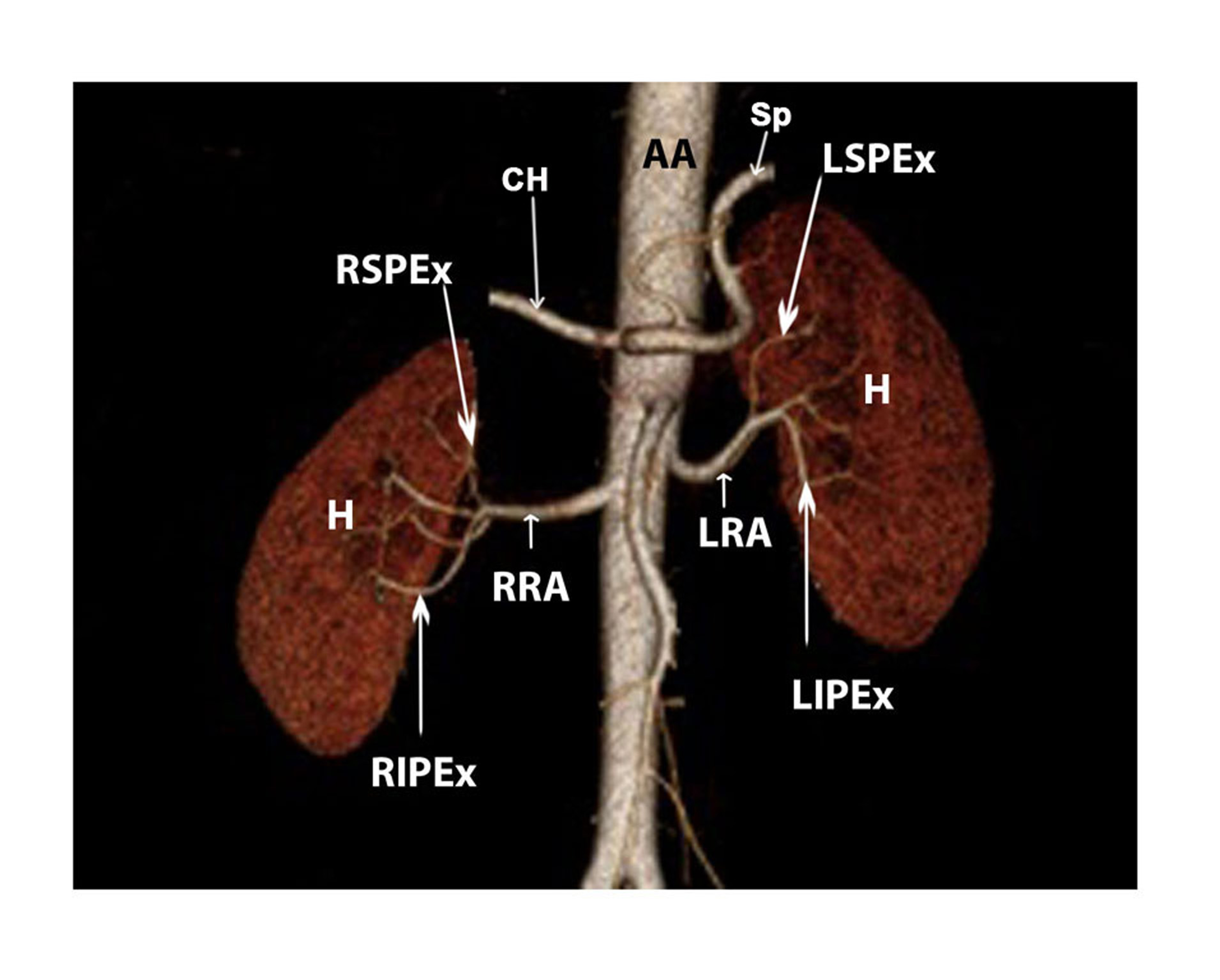
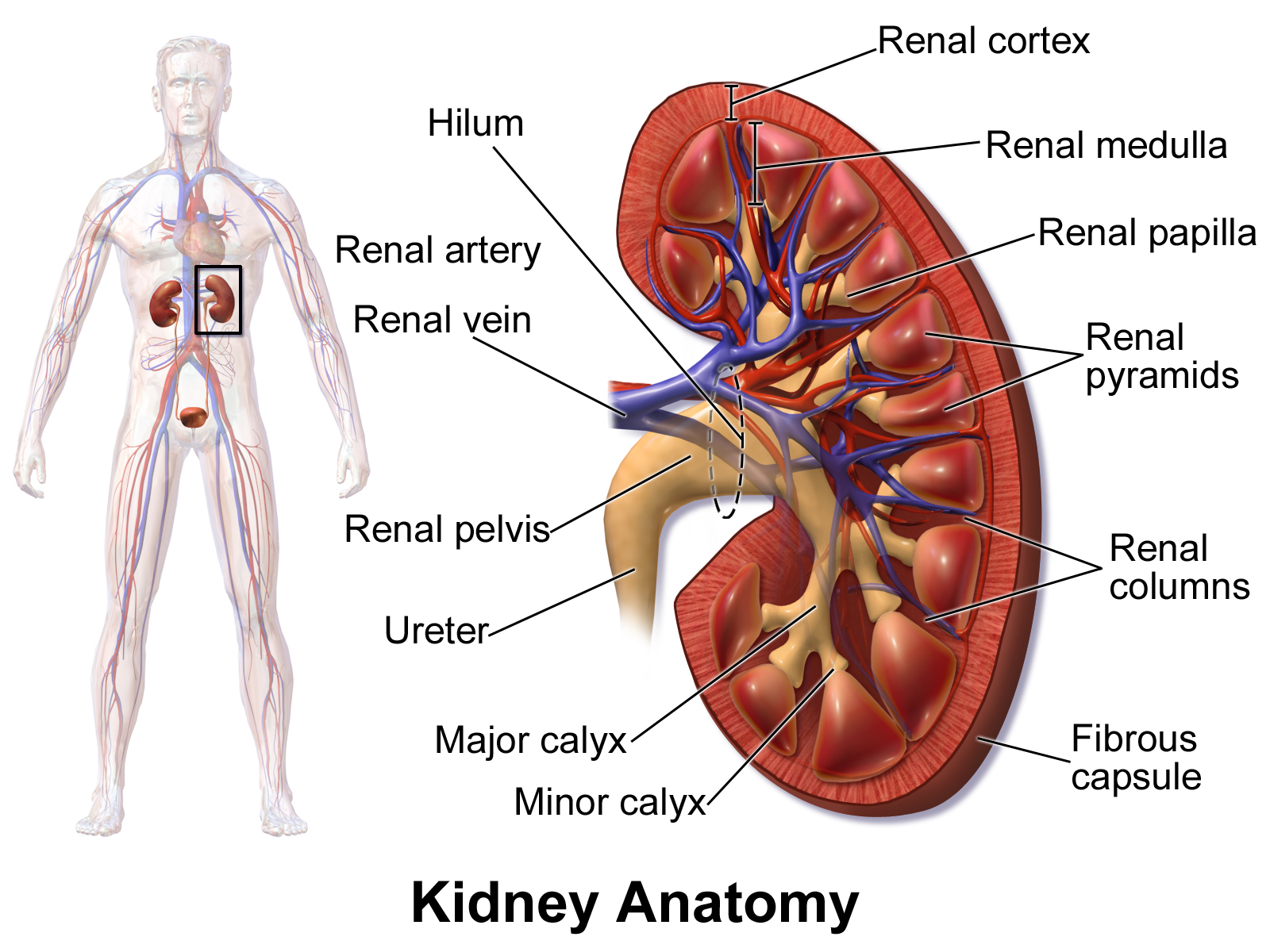
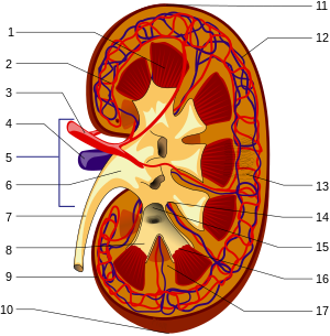
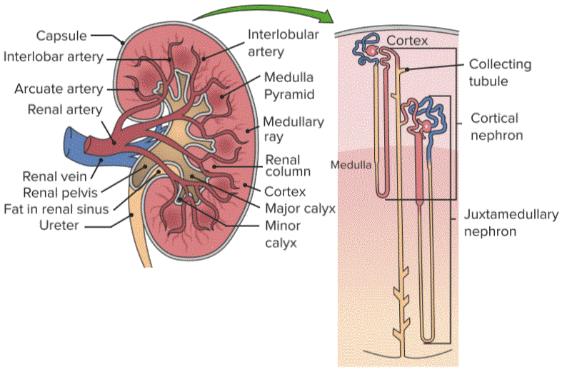
40 upper pole of kidney diagram
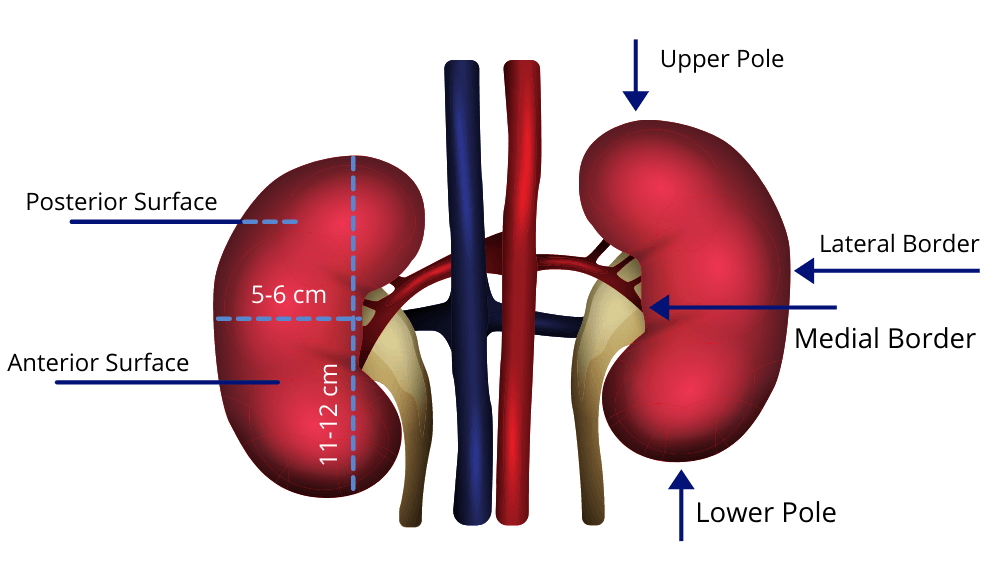
Upper Pole Nephrectomy. In some cases, the upper half of the kidney does not function from a ureterocele. If there is no urine reflux in the second ureter, the damaged part of the kidney may be removed. Often, this operation is done either through a small cut under the ribs, or laparoscopically. Nephrectomy Kidney Structure. The kidneys have a superior and inferior pole, medial and lateral margins, and an anterior and posterior surface. The superior pole of each kidney is deep to the rib cage. For the right kidney, its superior pole is at the 12th rib and for the left, the superior pole is at ribs 11 and 12.
Front of the left kidney is the tail of the pancreas, adjacent to the top of the spleen. In addition, the upper end (pole) of each kidney is in contact with the adrenal gland. Buds are covered in front by peritoneum. In the kidney is isolated front and rear surfaces, upper (extremitas superior) and lower (extremitas inferior) poles, or ends.

Upper pole of kidney diagram
ectopia type with upper pole of ectopic kidney fusing with lower pole of normal kidney. B- Sigmoid or S-shaped kidney where hilum of ectopic kidney faces laterally and that of normal kidney medially and with fusion form S-shaped mass. C- Lump kidney with fusion of two kidneys over a wide margin with ureter from ectopic kidney crossing the midline. The kidneys are two reddish-brown bean-shaped organs found in vertebrates. They are located on the left and right in the retroperitoneal space, and in adult humans are about 12 centimetres in length. They receive blood from the paired renal arteries; blood exits into the paired renal veins. Each kidney is attached to a ureter, a tube that carries excreted urine to the bladder. The kidney participates in the control of the volume of various body fluids, fluid osmolality, acid–base balance ... Diagram of Anterior anatomical relations of both kidneys. The kidneys are retroperitoneal organs that are located in the perirenal retroperitoneal space with a longitudinal diameter of 10–12 cm and a latero-lateral diameter of 3–5 cm and a weight of 250–270 g. In the supine position, the medial border of the normal kidney is much more anterior than the lateral border, The upper pole of each kidney is situated more posteriorly than the lower pole.
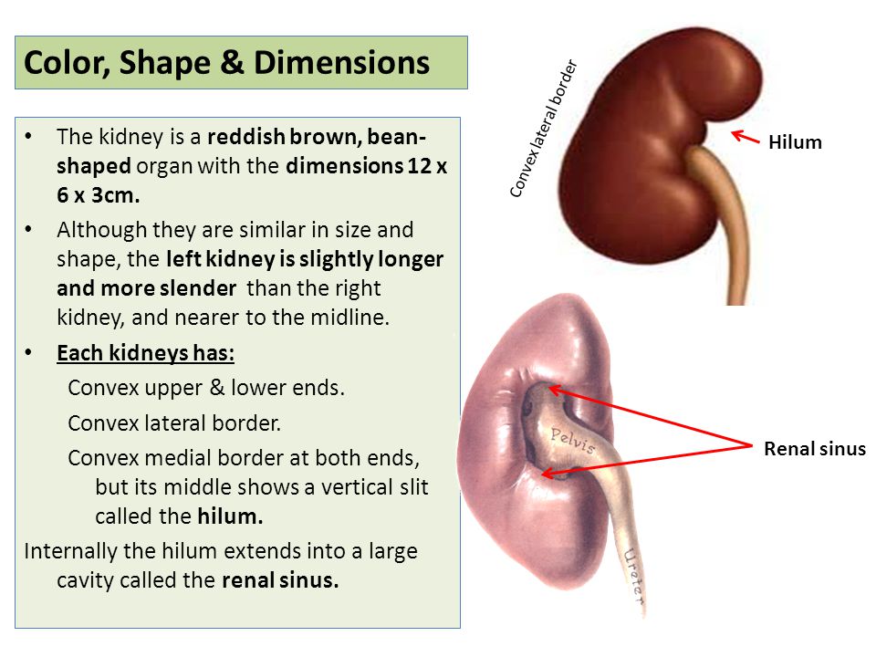
Upper pole of kidney diagram. Kidney Pain Location and Sensation. There is usually a feeling of a stabbing pain in the upper back just below the ribs. It may also be a dull ache depending on the diagnosis. At times kidney pain may be felt in the upper abdominal area. In this case it is often confused for digestive issues. Horseshoe kidney is the most common form of renal fusion. 64-66 It is the midline fusion of two distinct renal masses, each with its own ureter and pelvis (Figs. 1.28 and 1.29).Horseshoe kidney is relatively common (1:400 to 1:2000) with a 2:1 male predominance. Horseshoe kidney is commonly seen as part of other anomalies such as trisomy 18 (25%), caudal dysplasia syndrome, and Zellweger ... In general, stones in the lower pole of the kidney are more difficult to treat than stones in other areas of the kidney. Because of gravity, they don't float away as easily. Treatment options are based on stone size. ESWL or ureteroscopy is recommended for stones that are smaller than 1 cm in size. PCNL is needed for larger stones, but larger ... Kidneys are dark brown in colour and are embedded in a mass of fat. On the upper end of each kidney suprarenal glands are situated like a cap. Each kidney is about 10 to 13 cm (4- 5 inches) in long, 6 cm. (2 ½ inches) wide and 3 cm. (1 ½ inch) in thickness. The average weight of adult kidney is about 150 gms. in males and 135 gms in females.
Kidney cysts can occasionally lead to complications, including: An infected cyst. A kidney cyst may become infected, causing fever and pain. A burst cyst. A kidney cyst that bursts causes severe pain in your back or side. Urine obstruction. A kidney cyst that obstructs the normal flow of urine may lead to swelling of the kidney (hydronephrosis). Aug 29, 2017 · Overview. The kidneys are paired retroperitoneal structures that are normally located between the transverse processes of T12-L3 vertebrae, with the left kidney typically somewhat more superior in position than the right. The upper poles are normally oriented more medially and posteriorly than the lower poles. Calcification and the Kidneys. Calcification is the abnormal accumulation of calcium salts in body tissue. This abnormal accumulation of calcium in the kidney is referred to as nephrocalcinosis ... Anatomical Position. The kidneys lie retroperitoneally (behind the peritoneum) in the abdomen, either side of the vertebral column.. They typically extend from T12 to L3, although the right kidney is often situated slightly lower due to the presence of the liver.Each kidney is approximately three vertebrae in length. The adrenal glands sit immediately superior to the kidneys within a separate ...
If a kidney lesion is a solid mass, particularly one that picks up blood and thus “enhances” on contrast CT, it is considered malignant until proven otherwise. In the era of CT scan however, masses are found at a much smaller size than ever before. Now, if a mass is small, less than 2 cm, up to 20-25% of such lesions may be benign. Now let’s pay attention to the borders of the kidneys.A bean-like structure like the kidney has two borders: medial and lateral. The lateral border is directed towards the periphery, while the medial border is the one directed towards the midline. The medial border of the kidney contains a very important landmark called the hilum of the kidney, which is the entry and exit point for the ... What is upper pole of kidney? Overview. The kidneys are paired retroperitoneal structures that are normally located between the transverse processes of T12-L3 vertebrae, with the left kidney typically somewhat more superior in position than the right. The upper poles are normally oriented more medially and posteriorly than the lower poles. Q: I recently found that I have a 3mm non-obstructing calculus in the upper to mid pole of the left kidney as well as a 7mm cyst in the mid pole of the left kidney. I have had pain, which I am assuming is from the stone, for over two months. I drink a lot of water and was told by an urologist that the stone is too small to do anything with.
Investigation for the anatomy of the lower pole of the kidney (angle between lower infundibulum and pelvis, length and diameter of the infundibulum and number and pattern distribution of calyces) was carried out using intravenous pyelogram (IVP) in 100 cases with urinary stone (study cases) and 400 persons with normal kidneys (control subjects).
The left kidney is located slightly more superior than the right kidney due to the larger size of the liver on the right side of the body. Unlike the other abdominal organs, the kidneys lie behind the peritoneum that lines the abdominal cavity and are thus considered to be retroperitoneal organs. The ribs and muscles of the back protect the ...
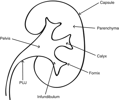
Case Discussion. Normally, as the kidney develops from lobules, each papilla has a corresponding calyx and infundibulum, emptying into the renal pelvis. In a compound calyx normal variant, multiple papillae empty into a single calyx and infundibulum. They are more common in the upper pole of the kidney. They are totally incidental.
mass upper pole right kidney. CountryLady46 posted: Recently I was sick with a bad sinus infection, had x-rays done, then had a CT scan done cause they saw a small spot on a lung. Well got results back on CT scan, lungs are fine BUT they did see a mass involving the upper pole of my right kidney. My question is: please name all the possible ...
Heterogeneous fat containing rounded exophytic mass extending off the upper pole of the right kidney measuring 11.7 x 10.7 x 15.4 cm. The angiomyolipoma demonstrates internal density consistent with hematoma and hematoma also seen in the right pararenal space extending particularly posterior and inferior to the kidney, the largest portion measuring approximately 8.7 x 3.2 x 6.6 cm.
Im 35 yrs of age and diagnose with septated cyst between the spleen and upper pole of the left kidney is it too bad and need to undergo operation? Dr. Robert Bennett answered. Urology 30 years experience. CYST: The size and presummed origin of the cyst determine the nature of the problem. A septation places it into a different category if it's ...
Learn term:kidney anatomy = upper, mid, and lower pole; with free interactive flashcards. Choose from 108 different sets of term:kidney anatomy = upper, mid, and lower pole; flashcards on Quizlet.
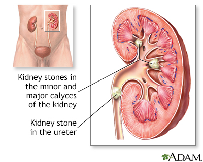
Urine contains many dissolved minerals and salts. When urine has high levels of minerals and salts, it can help to form stones. Kidney stones can start small but can grow larger in size, even filling the inner hollow structures of the kidney. Some stones stay in the kidney, and do not cause any problems. Sometimes, the kidney stone can travel down the ureter, the tube between the kidney and ...
A bilateral duplex collecting system is an unusual renal tract abnormality. Vesicoureteral reflux may be associated. We describe a rare case of bilateral duplex collecting system with bilateral vesicoureteral reflux in which the refluxing ureter on the left side drains the upper pole moiety contrary to what is often found. A 24-year-old married Arab woman presented with ascending left-sided ...
Feb 10, 2012 · 429 Background: Partial nephrectomy in upper pole kidney tumors represents a distinct surgical challenge. Data on minimally invasive nephron sparing surgery in this context are scarce. We set out to investigate the role of laparoscopic and open approaches to partial nephrectomy in these tumors. Methods: The Roswell Park Cancer Institute prospective, IRB-approved kidney surgery database was ...
The kidneys are retroperitoneal organs that are located in the perirenal retroperitoneal space with a longitudinal diameter of 10-12 cm and a latero-lateral diameter of 3-5 cm and a weight of 250-270 g. In the supine position, the medial border of the normal kidney is much more anterior than the lateral border, The upper pole of each kidney is situated more posteriorly than the lower pole.
Anatomy of the Kidney & Ureter. Paired Organ: Yes.. Each kidney or ureter is considered a separate primary, unless bilateral involvement is stated to be metastatic from one side to the other (exception: bilateral Wilms tumor of the kidney).. The kidneys have two functional areas that are managed and staged independently, the kidney parenchyma and the renal pelvis.
Adrenal gland are paired and located near the upper pole of kidney embedding in adipose tissue. Hi anatomy learner, if you are looking for the best guide to learn adrenal gland histology with different labeled slide and diagram then this article is for you. Fine, in this article I am going to share adrenal glands cortex and medulla histology in ...
What Size Kidney Cyst is Considered Large, and "When is a Kidney Cyst Considered Large". Usually depends on the size of the kidneys. For healthy adults, the kidneys are 10-12 cm long, 5-6 cm wide, and 3-4 cm thick. Small ganglion cysts usually do not cause annoying symptoms or complaints.
A sigle calculus is seen overlying the lower pole of left kidney that measures 4 mm in size. At least two (2) calculi are seen in the right kidney, one in the upper pole and one in the lower pole. A few diffuse smaller fragments are seen in the region of the right renal pelvis. A few phleboliths are seen in the pelvis. No calculi are seen along ...
Diagram of Anterior anatomical relations of both kidneys. The kidneys are retroperitoneal organs that are located in the perirenal retroperitoneal space with a longitudinal diameter of 10–12 cm and a latero-lateral diameter of 3–5 cm and a weight of 250–270 g. In the supine position, the medial border of the normal kidney is much more anterior than the lateral border, The upper pole of each kidney is situated more posteriorly than the lower pole.
The kidneys are two reddish-brown bean-shaped organs found in vertebrates. They are located on the left and right in the retroperitoneal space, and in adult humans are about 12 centimetres in length. They receive blood from the paired renal arteries; blood exits into the paired renal veins. Each kidney is attached to a ureter, a tube that carries excreted urine to the bladder. The kidney participates in the control of the volume of various body fluids, fluid osmolality, acid–base balance ...
ectopia type with upper pole of ectopic kidney fusing with lower pole of normal kidney. B- Sigmoid or S-shaped kidney where hilum of ectopic kidney faces laterally and that of normal kidney medially and with fusion form S-shaped mass. C- Lump kidney with fusion of two kidneys over a wide margin with ureter from ectopic kidney crossing the midline.

:watermark(/images/watermark_only.png,0,0,0):watermark(/images/logo_url.png,-10,-10,0):format(jpeg)/images/anatomy_term/left-renal-vein/y2EGhtsPklW5vKKTGuQ8jg_V._renalis_sinistra_02.png)







:background_color(FFFFFF):format(jpeg)/images/library/10926/renal-arteries_englishAA.jpg)


0 Response to "40 upper pole of kidney diagram"
Post a Comment