40 diagram of sinus cavity
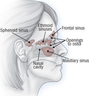
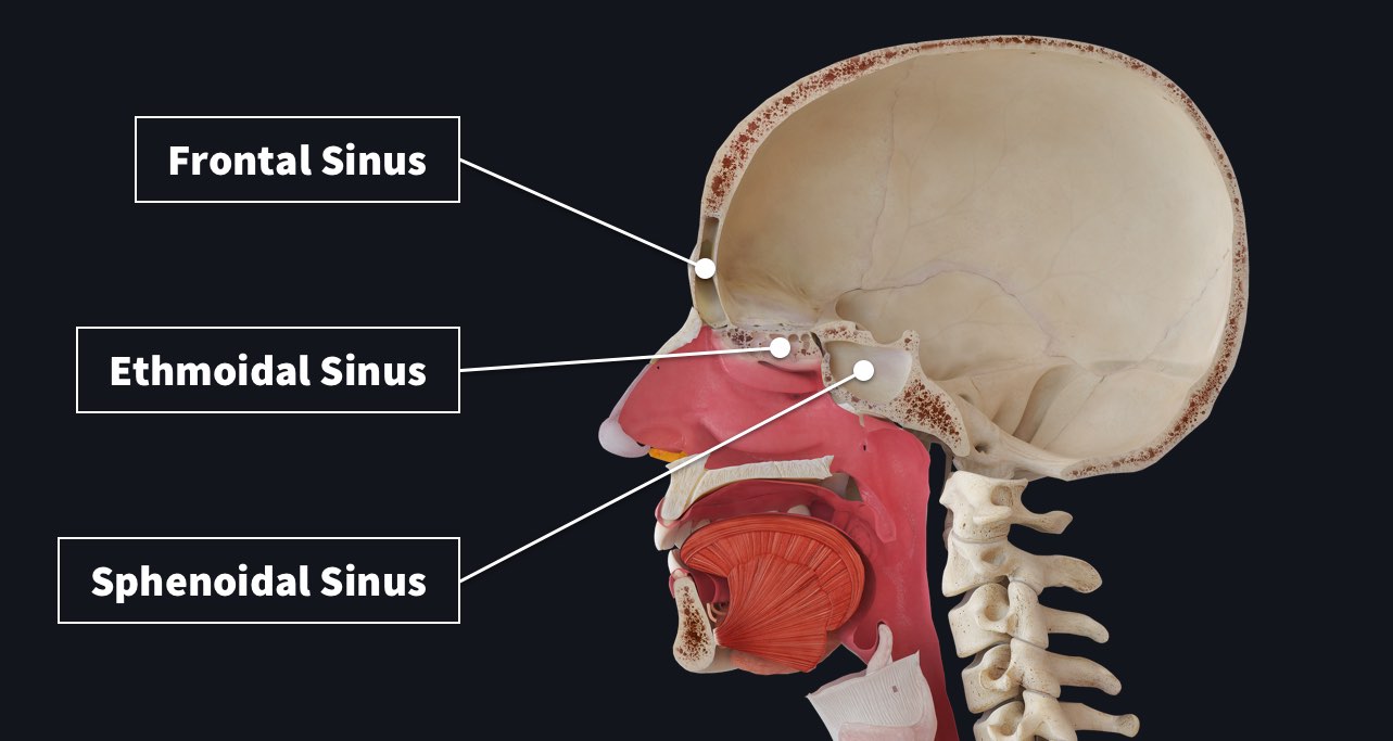
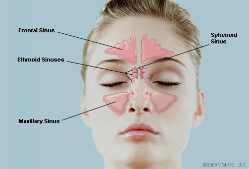
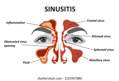
The sinuses are an air-filled cavity in a dense portion of a skull bone. They actually decrease the weight of the skull. The sinuses are formed in four right-left pairs. The frontal sinuses are positioned behind the forehead, while the maxillary sinuses are behind the cheeks. The sphenoid and ethmoid sinuses are deeper in the skull behind the ... Where do I get my information from: http://armandoh.org/resourceFacebook:https://www.facebook.com/ArmandoHasudunganSupport me: http://www.patreon.com/armando...
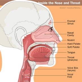
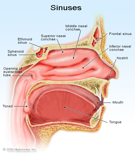
The nasal cavity consists of all the bones, tissues, blood vessels and nerves that make up the interior portion of the nose. The most important functions of the nasal cavity include warming and humidifying the air as you breathe and acting as a barrier for the immune system to keep harmful microbes from entering the body.

Diagram of sinus cavity
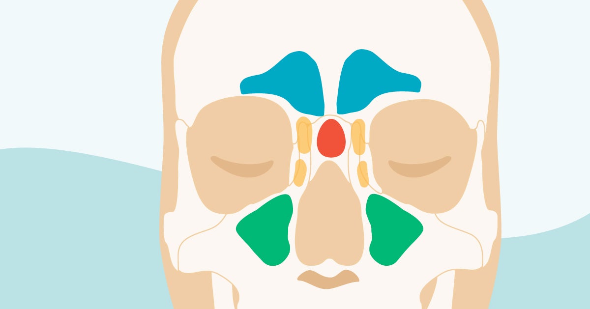
Maxillary Sinus (within the maxillary bones): The largest among all the paranasal sinuses [2], these two conical cavities are located on the two sides of the nose, above the upper teeth, and below the cheeks [4]. Ethmoid Sinus (within the ethmoid bones): Three to eighteen [5] air cells present in the ethmoid labyrinth, on both sides of the nose, between the eyes [6, 7]. Human body organ diagram front view. The dorsal body cavity protects organs of the nervous system and has two subdivisions. The cranial cavity is the area within the skull and encloses the brain. The medullary cavity contains red bone marrow during childhood eventually turning into yellow bone marrow after puberty. Inner body june 29 2019. Nasal Cavity Definition. The nose is one of the primary sensory organs responsible for the sense of smell, while it also plays major roles in respiration and speech production [1].The nasal cavity lies just behind the two nostrils and forms the interiors of the nose.. It makes up the upper respiratory system along with the paranasal sinuses, oral cavity, pharynx, and larynx [2], and is the ...
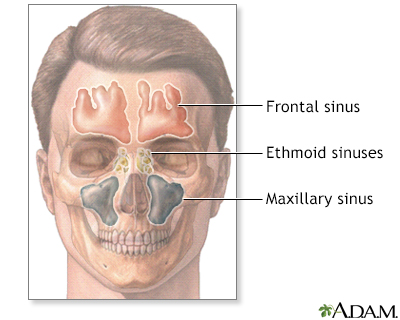
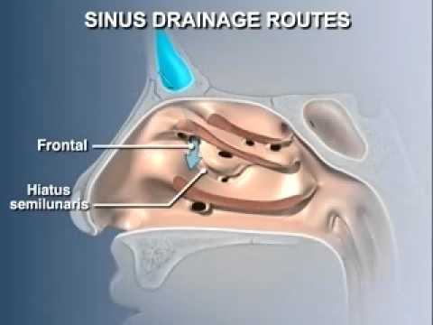
Diagram of sinus cavity. Diagram of Sinus Cavities and Drainage Ports What is Sinusitis? Sinusitis is caused by an inflammation of your sinus cavities that causes redness, swelling, mucus, and pain. There are two types of sinusitis: Acute sinusitis - an infection that is often triggered by the flu or cold. The flu or cold virus attacks your sinuses causing them to ... Sinus Pressure Points for Sinus Relief. Below are a number of sinus trigger points to help alleviate the discomfort from sinus pressure; some will even help mucus drain. A sinus pressure points diagram is also included to assist you in finding the location accurately on your face: The maxillary sinuses are the pockets near the cheeks. This sinus is located in the maxilla bone under the eye, contains three recesses, and is shaped like a pyramid. The frontal sinuses are over the forehead and above the eyes. These are absent at birth, and in approximately 5% of the adult population. The ethmoid sinuses are actually several ... View nasal cavity diagram videos. Browse 35 nasal cavity diagram stock illustrations and vector graphics available royalty-free, or start a new search to explore more great stock images and vector art. antique illustration: head throat section - nasal cavity diagram stock illustrations. cutaway diagram of human respiratory system, including ...
Browse 37 nasal cavity diagram stock photos and images available, or start a new search to explore more stock photos and images. antique illustration: head throat section - nasal cavity diagram stock illustrations. cutaway diagram of human respiratory system, including nasal and mouth cross section. - nasal cavity diagram stock illustrations. Neti Pot Diagram Of Sinus. WebMD examines the use of Neti pots to help relieve sinus problems and allergy symptoms. A neti pot a device used for nasal irrigation, and it looks like a little genie's lamp. ( Cute for something you stick in your nostril.) Its origins lie in. Nasal irrigation is a personal hygiene practice in which the nasal cavity ... The entire system is surrounded by a system of hollow cavities in the skull called the sinuses. The sinuses are about an inch across and are located in the cheekbones, the center of the forehead, between the eyes and in the nose. This interconnected ENT system helps us to breathe, smell and taste and plays a defining role in our looks. Diagrams of Sinus Cavities What is Sinusitis? Sinusitis is caused by an inflammation of your sinus cavities that causes redness, swelling, mucus, and pain. There are two types of sinusitis: Acute sinusitis - an infection that is often triggered by the flu or cold. The flu or cold virus attacks your sinuses causing them to swell and become narrow.
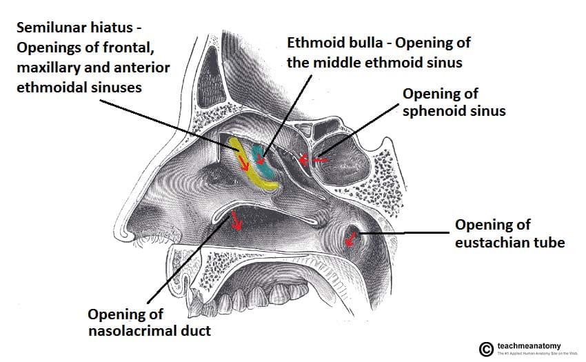
Nasal steroid spray: These medications ease tissue swelling and help prevent the regrowth of nasal polyps after sinus surgery. Nasal washes: They rinse mucus from the nasal cavities and sinuses. Jan 27, 2015 · Like the nasal cavity, the sinuses are all lined with mucus. The mucus secretions produced in the sinuses are continually being swept into the nose by the hair-like structures on the surface of ... The paranasal sinuses are air-filled extensions of the nasal cavity. There are four paired sinuses - named according to the bone in which they are located - maxillary, frontal, sphenoid and ethmoid. Each sinus is lined by a ciliated pseudostratified epithelium, interspersed with mucus-secreting goblet cells. Aug 10, 2011 · 1. SINUS CAVITIES DIAGRAM by Sinus Help www.sinusinfectionrelief.com 2. Fundamentally situated in the skull, human sinus cavities are the passageways mainly found in the areas around the face. Also known as paranasal sinuses, they are hollow, irregular air cavities which sit adjacent to and are attached to the nose and nasal passageways.
Anatomy Of The Human Nose Nasal Cavity Diagram Nose Text Hand People Png Pngwing
Aug 09, 2011 · sinus cavities diagram Published on Aug 9, 2011 Fundamentally situated in the skull, human sinus cavities are the passageways mainly found in the areas around the face.
The Paranasal Sinuses Structure Function Teachmeanatomy
Sinus Cavities Diagram. sinus cavities in the head anatomy diagram & sinuses often infections sinusitis is inflammation of a sinus caused by a bacterial infection that can follow a viral infection this causes pus and mucus to accumulate in the sinus symptoms can include fever headache stuffy nose and impaired sense of smell diagram sinus cavity anatomy diagram diagram sinus cavity see more ...

Diagram Of Sinus Cavity Sinus Cavities Paranasal Sinuses Anatomy And Physiology
The cavernous sinuses are 1 cm wide cavities that extend a distance of 2 cm from the most posterior aspect of the orbit to the petrous part of the temporal bone.They are bilaterally paired collections of venous plexuses that sit on either side of the sphenoid bone.Although they are not truly trabeculated cavities like the corpora cavernosa of the penis, the numerous plexuses, however, give the ...
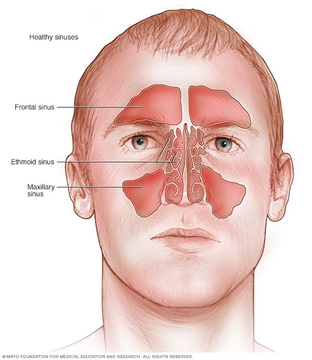
Healthy Sinuses Mayo Clinic
A CBCT scan of the sinuses shows a patient's paranasal sinus cavities. Paranasal sinuses are a group of four paired air-filled spaces that surround the nasal cavity. With a CBCT scanner, our allergists are able to see the sinuses located under the eyes, above the eyes, between the eyes, and behind the eyes.

425 Nasal Cavity Illustrations Clip Art Istock
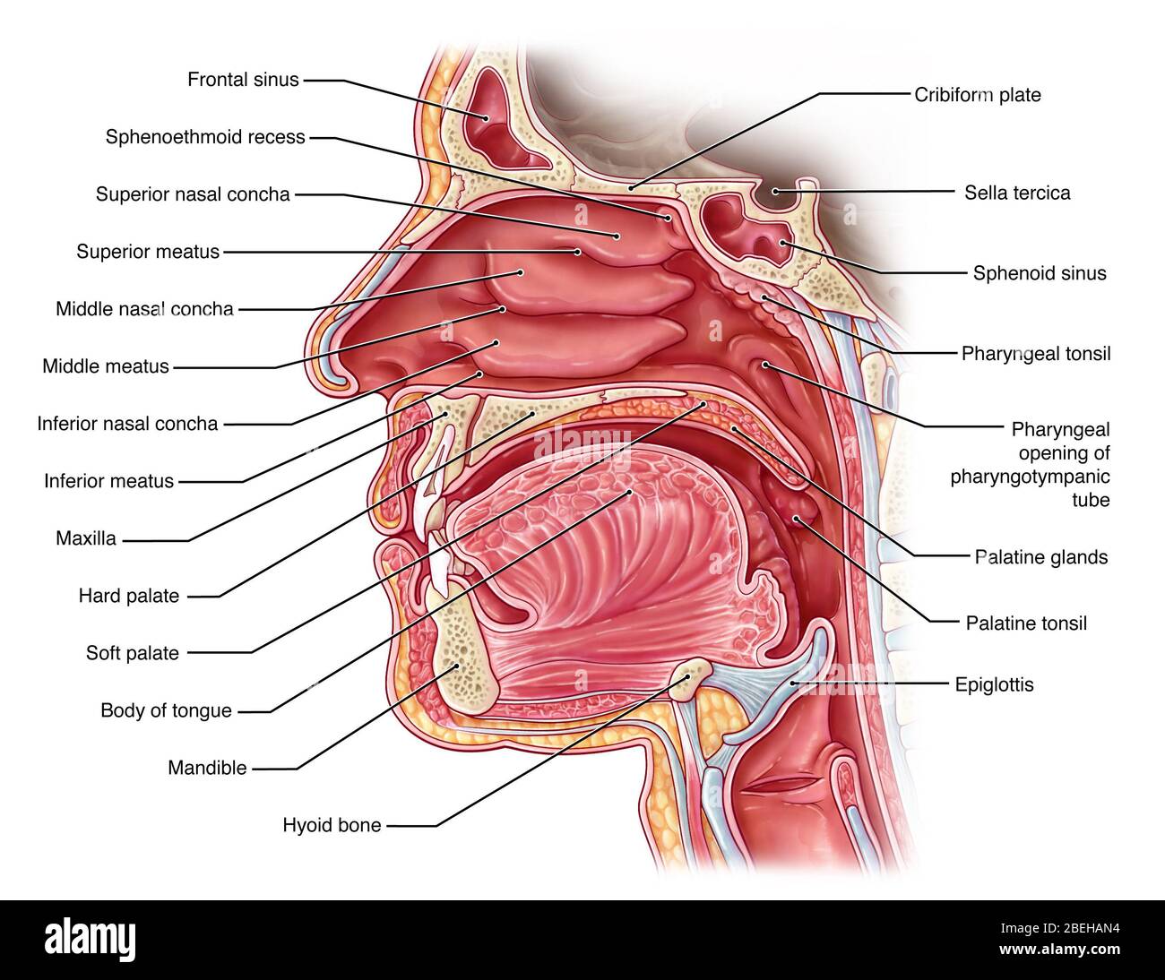
Besides the anterior and posterior apertures, each nasal cavity has a roof, floor, and lateral and medial walls.There are 12 cranial bones in total that contribute to the nasal cavity structure, which include the paired nasal, maxilla, palatine and lacrimal bones, as well as the unpaired ethmoid, sphenoid, frontal and vomer bones.Among all of them, the ethmoid bone is the most important ...

Anatomy Of The Nose And Paranasal Sinuses Osmosis
Aug 12, 2019 · There are four pairs of sinuses (named for the skull bones in which they're located). Interactive diagrams show sinus cavity locations and help visualize sinusitis, the most common type of sinus ...
Sinus Cavity High Resolution Stock Photography And Images Alamy
Sinuses of Nose. Human Anatomy - Sinus Diagram. Anatomy of the Nose. Nasal cavity bones. Anatomy of paranasal sinuses. Sinusitis - It is the inflammation of

Halotherapy And Your Sinus Halo Room
The nose is an olfactory and respiratory organ. It consists of nasal skeleton, which houses the nasal cavity. The nasal cavity has four functions: Warms and humidifies the inspired air.; Removes and traps pathogens and particulate matter from the inspired air. Responsible for sense of smell. Drains and clears the paranasal sinuses and lacrimal ducts.

Pin On Anatomy Upper Respiratory Tract
Hey, don't forget to check the frequently asked question and labelled diagram sections on animal body cavities with their contents. Structure of the animal nasal cavity. Each nasal cavity of an animal is filled largely by the ventral nasal conchae rostrally and ethmoturbinates caudally.

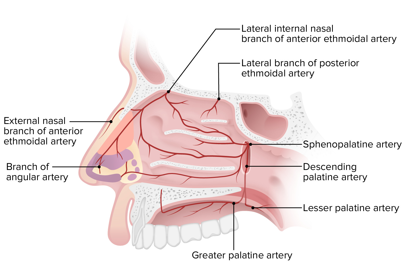
Figure Nasal Cavity Vasculature Used With Permission From Greys Anatomy Statpearls Ncbi Bookshelf
The space inside the nose is called the nasal cavity. This space warms, moistens and filters air as you breathe in. The bones around the nasal cavity have small hollow spaces in them called paranasal sinuses. These sinuses affect the sound and tone of your voice. Cancer that starts inside the nose or paranasal sinuses is called nasal and sinus ...
Nasal Cavity Radiology Reference Article Radiopaedia Org
A sinus is a sac or cavity in any organ or tissue, or an abnormal cavity or passage caused by the destruction of tissue.In common usage, "sinus" usually refers to the paranasal sinuses, which are air cavities in the cranial bones, especially those near the nose and connecting to it. Most individuals have four paired cavities located in the cranial bone or skull.
1 Nasal Cavity Anatomy Adapted From The Gray S Anatomy Of The Human Download Scientific Diagram
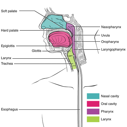
Nasal Cavity Definition. The nose is one of the primary sensory organs responsible for the sense of smell, while it also plays major roles in respiration and speech production [1].The nasal cavity lies just behind the two nostrils and forms the interiors of the nose.. It makes up the upper respiratory system along with the paranasal sinuses, oral cavity, pharynx, and larynx [2], and is the ...

Nasal Cavity Vector Illustration Of Human Nose Diagram Inside Of Nose Royalty Free Cliparts Vectors And Stock Illustration Image 66990051
Human body organ diagram front view. The dorsal body cavity protects organs of the nervous system and has two subdivisions. The cranial cavity is the area within the skull and encloses the brain. The medullary cavity contains red bone marrow during childhood eventually turning into yellow bone marrow after puberty. Inner body june 29 2019.
Anatomy And The Human Blockhead Anatomy Of The Nasal Cavity Howstuffworks
Maxillary Sinus (within the maxillary bones): The largest among all the paranasal sinuses [2], these two conical cavities are located on the two sides of the nose, above the upper teeth, and below the cheeks [4]. Ethmoid Sinus (within the ethmoid bones): Three to eighteen [5] air cells present in the ethmoid labyrinth, on both sides of the nose, between the eyes [6, 7].
Acute Sinusitis Harvard Health

Nasal Sinus Cavities Dalton Ga Aog Dalton

Premium Vector Human Sinus Cavity Diagram

1 Nasal Cavity Anatomy Adapted From The Gray S Anatomy Of The Human Download Scientific Diagram
Paranasal Sinuses Complete Anatomy

Sinuses Nasal Allergies
Sinusitis Information Mount Sinai New York
What Are The Sinuses Pictures Of Nasal Cavities

Nasal And Paranasal Sinus Anatomy And Embryology Ento Key

Postoperative Endoscopic Appearance Of Maxillary Sinus Cavities A Download Scientific Diagram
Anatomy Of The Nose Concise Medical Knowledge

Figure Anatomy Of The Paranasal Sinuses Spaces Between The Bones Around The Nose Pdq Cancer Information Summaries Ncbi Bookshelf

Nasal And Paranasal Sinus Anatomy And Embryology Ento Key
1
/GettyImages-165726373-178c670c99b44324836892cab60c5ac6.jpg)
The Nasal Cavity Anatomy Function And Treatment
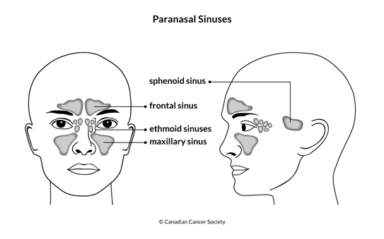
The Nasal Cavity And Paranasal Sinuses Canadian Cancer Society

9 Best Nasal Cavity Ideas Nasal Cavity Anatomy Anatomy And Physiology

Premium Vector Human Sinus Cavity Diagram
Anatomy Of The Nasal Cavity Mov Youtube

Pin By Dr Bikramaditya On Nasal Anatomy Biology Lessons Physiology Paranasal Sinuses

Topography Of The Skull Nasal Cavity

Normal Sinus Anatomy Medivisuals Medical Illustration Medical Illustration Sinusitis Anatomy
Sinus Cavity Images Stock Photos Vectors Shutterstock
Lippy Surgery Center Warren Oh Sinus And Nasal Surgery
Sinus Cavities In The Head Anatomy Diagram Pictures

Nose Sinus Cancer Anatomy
0 Response to "40 diagram of sinus cavity"
Post a Comment