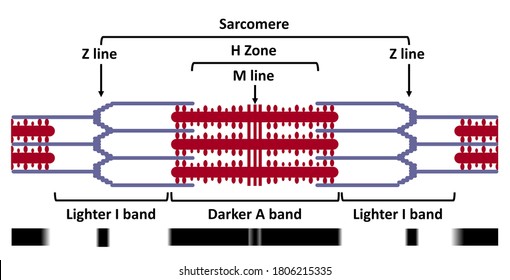
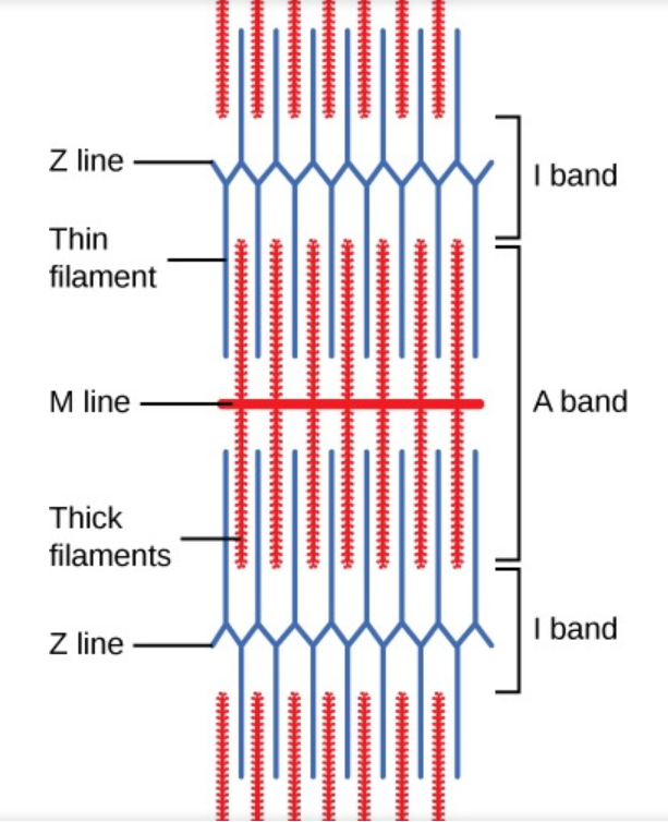
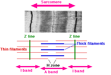
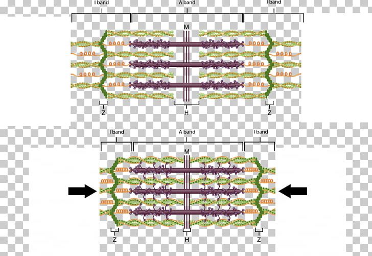
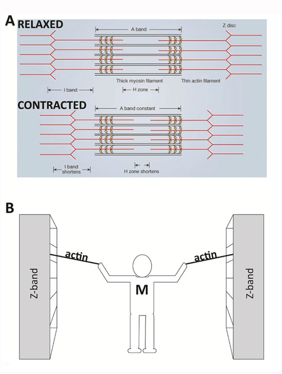
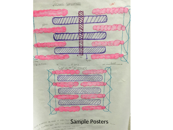
37 diagram of a sarcomere
Q 4. Draw the diagram of a sarcomere of skeletal muscle showing different regions. Biology. Q 5. Write true or false. If false change the statement so that it is true. (a) Actin is present in the thin filament. (b) H-zone of striated muscle fibre represents both thick and thin filaments. (b) Schematic diagram of a cardiac sarcomere. The sarcomere is the fundamental unit of contraction and is defined as the region between two Z-lines. Each ...
Sarcomere definition. A sarcomere is the functional unit of striated muscle. This means it is the most basic unit that makes up our skeletal muscle. Skeletal muscle is the muscle type that initiates all of our voluntary movement. Herein lies the sarcomere’s main purpose. Sarcomeres are able to initiate large, sweeping movement by contracting ...
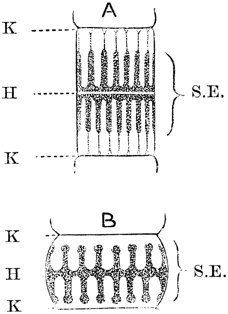
Diagram of a sarcomere
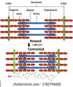
A sarcomere is the smallest functional unit of striated muscle tissue. It is the repeating unit between two Z-lines. Skeletal muscles are composed of ...Part of: Striated muscleLatin: sarcomerumBands · Contraction · Rest Draw a schematic diagram of a sarcomere contracting showing each of the 4 steps. close. Start your trial now! First week only $4.99! arrow_forward. Question. Draw a schematic diagram of a sarcomere contracting showing each of the 4 steps. check_circle Expert Answer. star. star. star. star. star. The thick filaments are anchored by a protein called myomesin at the center of the sarcomere (the M-line). The lighter I band regions contain thin actin ...1 answer · Top answer: Hint: Sarcomere is the essential unit of striated tissue in the muscles. This means that it is the most important entity that makes up our skeletal muscle. ...
Diagram of a sarcomere. Biology questions and answers. PART C: 7 questions total of 25 marks 1. The diagram below shows a sarcomere from a myofibril of a skeletal muscle. a) Label the diagram below to show all components of the sarcomere: A and I bands, H zone, M line. (2 marks) OOC OOO i. Why does muscle that has been overstretched produce less tension? (1 mark) ill. Z = last letter (last part of sarcomere or the border) Since the myosin and actin are sliding next to each other, all of those bands are going to change. The Z Line is just the border, so it won't change. 1. Share. Report Save. More posts from the Mcat community. 455. Posted by 7 days ago. (Refer to exam q) The diagram shows the stages in one cycle that results in movement of an actin filament in a muscle sarcomere. Describe how stimulation of a muscle by a nerve impulse starts the cycle shown in the diagram. (3) 2:17Presented by www.shikshaabhiyan.com This video is a part of the series for CBSE Class 11, Biology demo ...30 Jul 2019 · Uploaded by Shiksha Abhiyan
Question: i need help with these two pages. i think the top two are correct. the diagram for the sarcomere model is the last picture is for the 1st page. This problem has been solved! See the answer See the answer See the answer done loading. Figure 1 below shows a sarcomere from a skeletal muscle. Figure 2 shows a cross section through this sarcomere as it would appear when seen on an electron micrograph. (a)Copy figure 1 and draw on a line to show from where the cross-section the muscle in figure 2 was taken. (b)Name the main protein found in region P. Each myofibril is made up of contractile sarcomeres AND Drawing labelled diagrams of the structure of a sarcomere. Draw the diagram of a sarcomere of skeletal muscle showing different regions. Answer. The diagrammatic representation of a sarcomere is as follows: ... -Describe various types of epithelial tissues with the help of labelled diagrams. Q:-What happens to the respiratory process in a man going up a hill? Q:-Define a cardiac cycle and the cardiac ...
The diagrams show a sarcomere in different states of contraction. a. Name the parts labelled P, Q and R. b. Explain why there are no actin–myosin cross-bridges visible in diagram A. c. Muscle fibres are able to contract with more force in some states of contraction than others. Suggest which of the diagrams shows the state that can develop ... a Schematic diagram showing the main components of the sarcomere. The A-band comprises myosin filaments crosslinked at the centre by the M-band assembly. Thin ... The thick filaments are anchored by a protein called myomesin at the center of the sarcomere (the M-line). The lighter I band regions contain thin actin ...1 answer · Top answer: Hint: Sarcomere is the essential unit of striated tissue in the muscles. This means that it is the most important entity that makes up our skeletal muscle. ... Draw a schematic diagram of a sarcomere contracting showing each of the 4 steps. close. Start your trial now! First week only $4.99! arrow_forward. Question. Draw a schematic diagram of a sarcomere contracting showing each of the 4 steps. check_circle Expert Answer. star. star. star. star. star.
A sarcomere is the smallest functional unit of striated muscle tissue. It is the repeating unit between two Z-lines. Skeletal muscles are composed of ...Part of: Striated muscleLatin: sarcomerumBands · Contraction · Rest
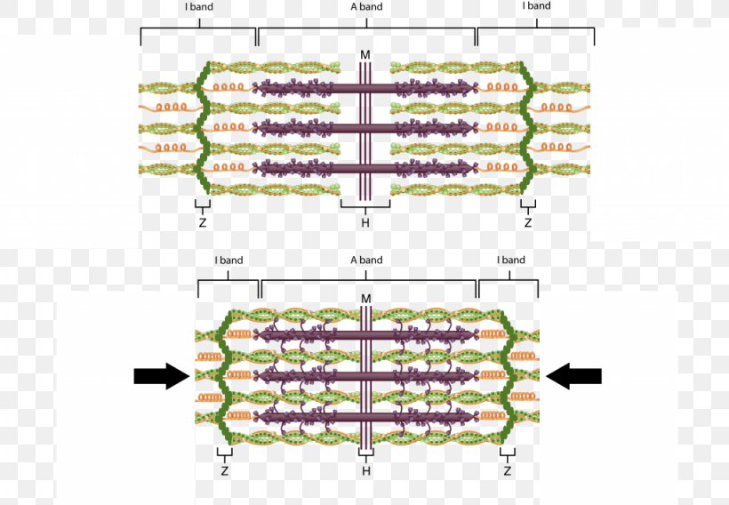
Diagram Sarcomere Myofilament Skeletal Muscle Muscle Contraction Png 1024x710px Diagram Actin Anatomy Area Motor Unit Download

The Sarcomere And Sliding Filaments In Muscular Contraction Definition And Structures Video Lesson Transcript Study Com
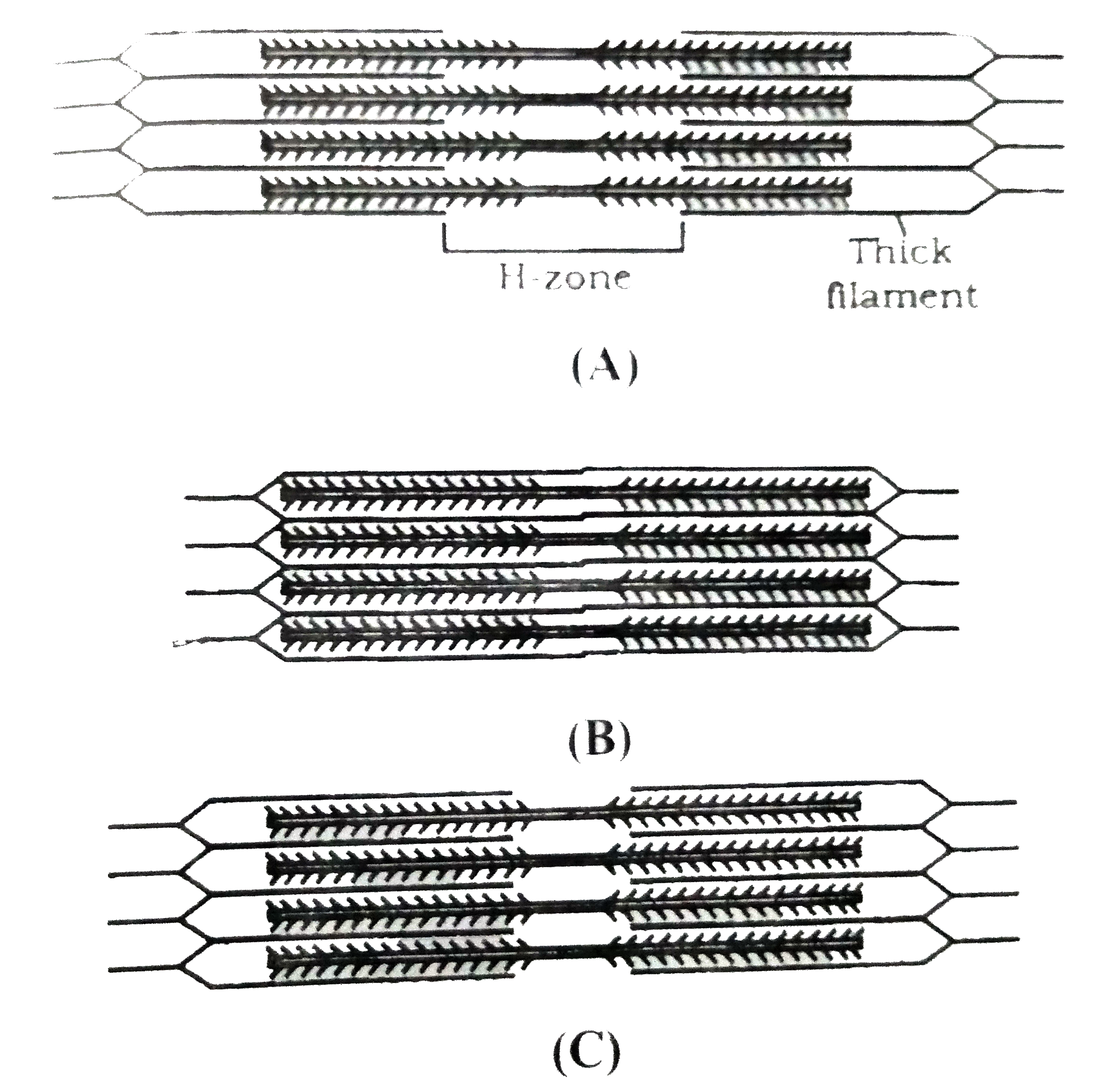
Look At The Given Diagram Of Events During Muscle Contraction Named As A B C Identidy The Diagram That Shows Gradual Shortening Of Length Of Sarcomere Img Src Https D10lpgp6xz60nq Cloudfront Net Physics Images Aln Bio C15 E39 001 Q01 Png
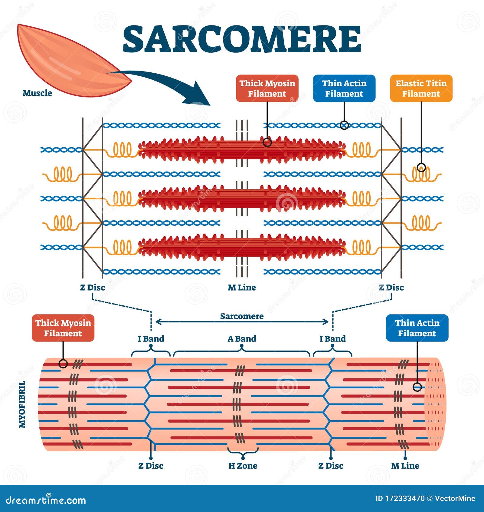
Sarcomere Muscular Biology Scheme Vector Illustration Stock Vector Illustration Of Contraction Diagram 172333470

Muscular System Skeletal Muscle Contraction Sarcomeres And The Sliding Filament Model Diagram Quizlet











0 Response to "37 diagram of a sarcomere"
Post a Comment